. 2013 Nov;17(11):1397-409.
doi: 10.1111/jcmm.12156. Epub 2013 Nov 19.

Combination treatment for myeloproliferative neoplasms using JAK and pan-class I PI3K inhibitors

Affiliations

Combination treatment for myeloproliferative neoplasms using JAK and pan-class I PI3K inhibitors

Meng Ling Choong et al. J Cell Mol Med. 2013 Nov.

Abstract

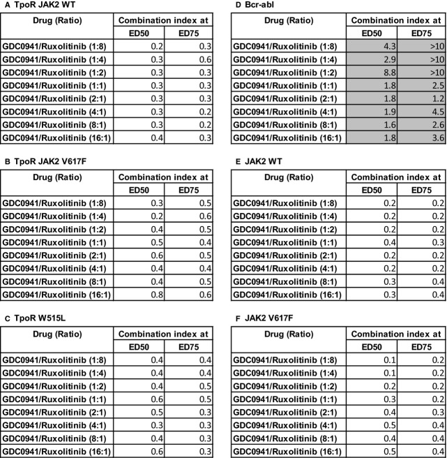
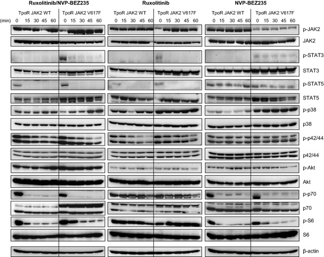
Current JAK2 inhibitors used for myeloproliferative neoplasms (MPN) treatment are not specific enough to selectively suppress aberrant JAK2 signalling and preserve physiological JAK2 signalling. We tested whether combining a JAK2 inhibitor with a series of serine threonine kinase inhibitors, targeting nine signalling pathways and already used in clinical trials, synergized in inhibiting growth of haematopoietic cells expressing mutant and wild-type forms of JAK2 (V617F) or thrombopoietin receptor (W515L). Out of 15 kinase inhibitors, the ZSTK474 phosphatydylinositol-3'-kinase (PI3K) inhibitor molecule showed strong synergic inhibition by Chou and Talalay analysis with JAK2 and JAK2/JAK1 inhibitors. Other pan-class I, but not gamma or delta specific PI3K inhibitors, also synergized with JAK2 inhibitors. Synergy was not observed in Bcr-Abl transformed cells. The best JAK2/JAK1 and PI3K inhibitor combination pair (ruxolitinib and GDC0941) reduces spleen weight in nude mice inoculated with Ba/F3 cells expressing TpoR and JAK2 V617F. It also exerted strong inhibitory effects on erythropoietin-independent erythroid colonies from MPN patients and JAK2 V617F knock-in mice, where at certain doses, a preferential inhibition of JAK2 V617F mutated progenitors was detected. Our data support the use of a combination of JAK2 and pan-class I PI3K inhibitors in the treatment of MPNs.

Keywords: JAK2; PI3K; combination treatment; kinases; myeloproliferative neoplasms.


Figures

Figure 1
Figure 1
Cell lines and small molecules used in combination for detection of synergy with JAK2 inhibitors in inhibiting proliferation of model myeloproliferative neoplasm cells. (A) Ba/F3 cell lines used for inhibitor screens. Ba/F3 parental and Ba/F3 TpoR JAK2 wild-type (WT) cells were maintained in medium supplemented with IL3, while Ba/F3 JAK2 V617F, TpoR V617F, TpoR W515L and Bcr-Abl were maintained in medium without cytokines. (B) A schematic representation of an 8 × 8 constant ratio design for combination treatment. The two combination drugs were used at their equipotent concentration ratio (IC50 of drug A to IC50 of drug B is 1:1) in the centre column. The concentration ratio of drug A to drug B is progressively increased towards the left, while the concentration ratio of drug B to drug A is progressively increased towards the right. Each column in the design is a dose–response curve with constant concentration ratio between drug A and drug B. Although usually C.I. <0.8 is considered significant, we selected for combinations showing C.I. <0.5. (C) Combination study using JAK2/JAK1 inhibitor ruxolitinib with various kinase inhibitors at equipotent concentration ratio on TpoR JAK2 V617F cells. (D) Combination study using JAK2/JAK1 inhibitor ruxolitinib with several other PI3K inhibitors at equipotent concentration ratio on TpoR JAK2 V617F cells.
Figure 2
Figure 2
Combination study using JAK2/JAK1 inhibitor ruxolitinib with ZSTK474 in the Ba/F3 cell model. (A) Ba/F3 TpoR JAK2 wild-type (WT) cells; (B) Ba/F3 TpoR JAK2 V617F cells; (C) Ba/F3 TpoR W515L cells; (D) Ba/F3 Bcr-Abl cells; (D and E) Ba/F3 JAK2 WT cells; (F) Ba/F3 JAK2 V617F cells.
Figure 3
Figure 3
Combination study using the JAK2/JAK1 inhibitor ruxolitinib with GDC0941in the Ba/F3 cell model. Results shown are for eight dose ratios and at two effective doses (ED50 and ED75). (A) Ba/F3 TpoR JAK2 wild-type (WT) cells; (B) Ba/F3 TpoR JAK2 V617F cells; (C) Ba/F3 TpoR W515L cells; (D) Ba/F3 Bcr-Abl cells; (D and E) Ba/F3 JAK2 WT cells; (F) Ba/F3 JAK2 V617F cells.
Figure 4
Figure 4
Effect of the JAK2/JAK1 inhibitor ruxolitinib and PI3K inhibitor NVP-BEZ236 alone or in combination on signalling in Ba/F3 TpoR cells overexpressing JAK2 wild-type (WT) or JAK2 V617F. Cells expressing TpoR JAK2 V617F were autonomous for growth, while Ba/F3 TpoR JAK2 WT cells were grown in medium supplemented with IL3. Cells were treated for the indicated intervals with 1 μM ruxolitinib and 10 μM NVP-BEZ235 alone or in combination and western blotting was performed with the phospho-specific antibodies (phospho-JAK2 (Tyr1007/1008), phospho-STAT3 (Tyr705), phospho-STAT5 (Tyr694), phospho-p70 S6 kinase (Thr389), phospho-S6 ribosomal protein (Ser235/236), phospho-AKT (Ser473), phospho-p38 MAP-kinase (Thr180/Tyr182), phospho-p44/42 ERK1/2 (Thr202/Tyr204), with antibodies to actin (as loading control) and with antibodies detecting the signalling proteins themselves.
Figure 5
Figure 5
Effect of JAK and PI3K inhibitions on survival (A) and spleen weight (B) in the JAK2 mutation-driven leukaemia mouse model. Ba/F3 TpoR JAK2 V617F cells (1 × 107) were intravenously inoculated into 8- to 10-week-old female nude mice. After 3 days, mice were treated with either vehicle, GDC0941 alone at 50 mg/kg body weight, ruxolitinib alone at 50 mg/kg body weight, or a combination of both compounds for 12 days. Combination treatment increased survival by 2 days compared with single compound treatment. Combination treatment also reduced spleen weight significantly compared with vehicle-treated mice, or mice treated with GDC0941 alone, or ruxolitinib alone (*P < 0.001 for each comparison). qd: every day, bid: twice a day.
Figure 6
Figure 6
Effect of JAK and PI3K inhibitors on colony formation from heterozygous JAK2 V617F knock-in bone marrow cells and mixtures of knock-in and wild-type (WT) mice. (A) Effect of ruxolitinib (5 μM) and GDC 0942 (10 μM) or combination thereof on Epo-independent CFU-E (2% foetal serum, no cytokines) from bone marrow of reconstituted heterozygous JAK2 V617F knock-in mice. The number of colonies was normalized to those in the untreated controls in the absence of Epo (936 ± 302 EEC/200,000 bone marrow nucleated heterozygous JAK2 V617F knock-in cells). Shown are averages of two mice, each performed in duplicate (average of six points) ±SD. **P < 0.01 unpaired Student's test with unequal variance. (B) Comparison between generation of CFU-E colonies between bone marrow cells from JAK2 V617F knock-in mice and JAK2 WT littermate mice. Cells were plated in methylcellulose in the presence of cytokines (3 U/ml Epo, 5 ng/ml SCF, 3 ng/ml IL3) alone or with the combination of 0.1 μM ruxolitinib and 1 μM GDC 0941. The number of colonies was normalized to those in the untreated controls in presence of cytokines, which was 1099 ± 56/150,000 bone marrow nucleated heterozygous JAK2 V617F cells and 837 ± 185/150,000 bone marrow nucleated WT JAK2 cells. Shown are averages of triplicates ±SD of one representative experiment out of two. *P < 0.05 unpaired Student's test with unequal variance. (C) Bone marrow cells from JAK2 V617F knock-in mice and WT littermate mice were mixed in a 1:1 ratio (75,000 cells each) and BFU-E formation was determined in the absence or presence of the combination of 0.1 μM ruxolitinib and 1 μM GDC 0941 in the presence of cytokines (3 U/ml Epo, 5 ng/ml SCF, 3 ng/ml IL3). The number of colonies was normalized to those in the untreated controls in the presence of cytokines, which was 339 ± 98 BFU-E colonies/150,000 nucleated cells. Shown are averages of triplicates ±SD of one representative experiment out of two. *P < 0.05 unpaired Student's test with unequal variance. (D) Preferential inhibition of JAK2 V617F knock-in BFU-E compared with WT BFU-E by the combination of 0.1 μM ruxolitinib and 1 μM GDC 0941. 1:1 mixed JAK2 V617F knock-in and WT cells were plated in methylcellulose in the presence of cytokines (3 U/ml Epo, 5 ng/ml SCF, 3 ng/ml IL3) alone or with the combination of inhibitors. BFU-E colonies were harvested at day 6 from three plates for each condition and genotyped for JAK2 V617F. Three plates were used for each condition. Genotyping was performed on 4, 2 and 5 BFU-Es for the condition ‘cytokines’ and 4, 6 and 6 for the condition ‘cytokines + inhibitor’ combination. Values represent% of BFU-Es genotyped as JAK2 V617F-positive or JAK2 WT-positive per 150,000 nucleated mixed cells. Shown are the results of one representative experiment out of two.

Similar articles

Cited by

References

    1. James C, Ugo V, Couedic Le JP, et al. A unique clonal JAK2 mutation leading to constitutive signalling causes polycythaemia vera. Nature. 2005;434:1144–8. - PubMed
    1. Kralovics R, Passamonti F, Buser AS, et al. A gain-of-function mutation of JAK2 in myeloproliferative disorders. N Engl J Med. 2005;352:1779–90. - PubMed
    1. Levine RL, Wadleigh M, Cools J, et al. Activating mutation in the tyrosine kinase JAK2 in polycythemia vera, essential thrombocythemia, and myeloid metaplasia with myelofibrosis. Cancer Cell. 2005;7:387–97. - PubMed
    1. Baxter EJ, Scott LM, Campbell PJ, et al. Acquired mutation of the tyrosine kinase JAK2 in human myeloproliferative disorders. Lancet. 2005;365:1054–61. - PubMed
    1. Levine RL, Pardanani A, Tefferi A, et al. Role of JAK2 in the pathogenesis and therapy of myeloproliferative disorders. Nat Rev Cancer. 2007;7:673–83. - PubMed

Publication types

MeSH terms