. 2014 Jan;18(1):69-79.
doi: 10.1111/jcmm.12165. Epub 2013 Nov 19.

Motor protein function in skeletal abdominal muscle of cachectic cancer patients

Affiliations

Motor protein function in skeletal abdominal muscle of cachectic cancer patients

Sultan Taskin et al. J Cell Mol Med. 2014 Jan.

Abstract

Cachexia presents with ongoing muscle wasting, altering quality of life in cancer patients. Cachexia is a limiting prognostic factor for patient survival and health care costs. Although animal models and human trials have shown mechanisms of motorprotein proteolysis, not much is known about intrinsic changes of muscle functionality in cancer patients suffering from muscle cachexia, and deeper insights into cachexia pathology in humans are needed. To address this question, rectus abdominis muscle samples were collected from several surgical control, non-cachectic and cachectic cancer patients and processed for skinned fibre biomechanics, molecular in vitro motility assays, myosin isoform protein compositions and quantitative ubiquitin polymer protein analysis. In pre-cachectic and cachectic cancer patient samples, maximum force was significantly compromised compared with controls, but showed an unexpected increase in myofibrillar Ca(2+) sensitivity consistent with a shift from slow to fast myosin isoform expression seen in SDS-PAGE analysis and in vitro motility assays. Force deficit was specific for 'cancer', but not linked to presence of cachexia. Interestingly, quantitative ubiquitin immunoassays revealed no major changes in static ubiquitin polymer protein profiles, whether cachexia was present or not and were shown to mirror profiles in control patients. Our study on muscle function in cachectic patients shows that abdominal wall skeletal muscle in cancer cachexia shows signs of weakness that can be partially attributed to intrinsic changes to contractile motorprotein function. On protein levels, static ubiquitin polymeric distributions were unaltered, pointing towards evenly up-regulated ubiquitin protein turnover with respect to ubiquitin conjugation, proteasome degradation and de-ubiquitination.

Keywords: cancer; contractility; motility assay; muscle cachexia; myosin isoforms.


Figures

Figure 1
Figure 1
Ca2+ sensitivity of the contractile apparatus and maximum force values from rectus abdominis muscle biopsies in control, non-cachectic and cachectic cancer patients. (A) Representative recording of force response to increasing Ca2+ concentrations (decreasing pCa) in one control patient sample (left panel; ordinate—relative force units; abscissa—time). Right panel shows the mean pCa-force curves reconstructed from group data and fitted with a sigmoidal fit. For a more quantitative analysis, steady-state force relations from each patient within a group were fitted with a Hill function and pCa50 and Hill coefficient extracted. (B) Mean values of pCa50 indicate a significantly increased Ca2+ sensitivity of the contractile apparatus in cachectic versus non-cachectic patients, while Hill coefficients h were similar. (C) Maximum absolute force was significantly compromised in samples from cancer patients, unrelated to the presence of cachexia. n/m indicates number of fibre bundles n studied in m participants within each group and are the same in (B) and (C). *P < 0.05.
Figure 2
Figure 2
In vitro motility assays suggest a shift of slow to intermediate sliding velocities in cancer patients. (A) Representative images from actin-filament sliding sequences of a control patient single cell extract. Two individual filaments are tracked in time (arrow, asterisk). Velocity distributions for each experiment were assessed and fitted using a Gaussian curve to extract median velocities (B). (C) Median sliding velocities for each patient group revealed a clear double-peak histogram distribution in control participants, but a triple-peak velocity distribution in non-cachectic cancer patients. A triple band for velocity was empirically defined according to maximized correlation coefficients of the Gaussian multi-peak fits. This gave a cut-off of <4.25 μm/sec. for slow velocities, intermediate velocities ranged between 4.25 μm/sec. and 6.25 μm/sec. and >6.25 μm/sec. for fast sliding velocities. (D) For each patient, percentage distribution of filament velocities in each velocity group shows substantial scattering (lower panel). Almost no intermediate filament velocities were found in control patients, while there was a tendency towards higher intermediate filament velocity percentages in cancer patients, mainly because of a reduction in slow filaments (upper panel).
Figure 3
Figure 3
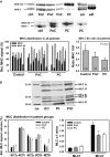
SDS-PAGE analysis of motorprotein distribution suggests a shift in heavy chain distribution towards faster isoforms in cachectic cancer patients. (A) Two MHC gels from rectus abdominis muscle samples from control (ctrl), non-cachectic (PnC) and cachectic cancer patients (PC) are shown with a sample from murine soleus (sol) and extensor digitorum longus (edl) muscles, for which isoform distribution is well known. Although there is some scattering between patients regarding relative contents of MHC-I and MHC-IIA signals, there is a consistently larger fraction of faster MHC-IIA in the PC group. This is also reflected by a largely reduced MHC I:IIA ratio, which declines from control to non-cachectic and cachectic patients. Myosin light chains MLC isoforms were analysed from gels such as shown in (B). Apart from a significant smaller fraction of MLC-2s over MLC-2f in cancer patients over controls, this was not significant in cachectic patients and there was no major difference in the other isoforms (*P < 0.05 versus control).
Figure 4
Figure 4
Quantitative ubiquitin Western blot analysis indicates similar static protein amounts of some ubiquitin multimer levels in abdominal muscle from control, non-cachectic and cachectic cancer patients. (A) Representative Ubiquitin Western blot of rectus abdominis muscle samples from control, non-cachectic and cachectic tumour patients (left panel), as well as relative signal volume densities from individual patients (right panel). Volume densities on their own suggest vastly enlarged dimer fractions and significantly reduced monomer fractions in tumour patients over controls, an observation which is no longer present when normalizing ubiquitin to GAPDH (B). With the slopes from the calibration curves for multimers (Fig. S1), multimer protein weights and their relative distribution were quantified (C). Patient identifiers in (A) are: #484 = ctrl#3; #486 = PnC#4; #481 = PC#2.

References

    1. Tisdale MJ. Mechanisms of cancer cachexia. Phys Rev. 2009;89:381–410. - PubMed
    1. Fortunati N, Manti R, Birocco N, et al. Pro-inflammatory cytokines and oxidative stress/antioxidant parameters characterize the bio-humoral profile of early cachexia in lung cancer patients. Oncol Rep. 2007;18:1521–7. - PubMed
    1. Cai D, Frantz JD, Tawa NE, Jr, et al. IKKbeta/NFkappaB activation causes severe muscle wasting in mice. Cell. 2004;119:285–98. - PubMed
    1. Bachmann J, Heiligensetzer M, Krakowski-Roosen H, et al. Cachexia worsens prognosis in patients with resectable pancreatic cancer. J Gastrointest Surg. 2008;12:1193–201. - PubMed
    1. Tisdale MJ. Cachexia in cancer patients. Nat Rev Cancer. 2002;2:862–71. - PubMed

Publication types

MeSH terms