Expression and distribution of GABA and GABAB-receptor in the rat adrenal gland
- PMID: 24252118
- PMCID: PMC3969063
- DOI: 10.1111/joa.12144
Expression and distribution of GABA and GABAB-receptor in the rat adrenal gland
Abstract
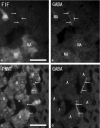
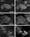
The inhibitory effects of gamma-aminobutyric acid (GABA) in the central and peripheral nervous systems and the endocrine system are mediated by two different GABA receptors: GABAA-receptor (GABAA-R) and GABAB-receptor (GABAB-R). GABAA-R, but not GABAB-R, has been observed in the rat adrenal gland, where GABA is known to be released. This study sought to determine whether both GABA and GABAB-R are present in the endocrine and neuronal elements of the rat adrenal gland, and to investigate whether GABAB-R may play a role in mediating the effects of GABA in secretory activity of these cells. GABA-immunoreactive nerve fibers were observed in the superficial cortex. Some GABA-immunoreactive nerve fibers were found to be associated with blood vessels. Double-immunostaining revealed GABA-immunoreactive nerve fibers in the cortex were choline acetyltransferase (ChAT)-immunonegative. Some GABA-immunoreactive nerve fibers ran through the cortex toward the medulla. In the medulla, GABA-immunoreactivity was seen in some large ganglion cells, but not in the chromaffin cells. Double-immunostaining also showed GABA-immunoreactive ganglion cells were nitric oxide synthase (NOS)-immunopositive. However, neither immunohistochemistry combined with fluorescent microscopy nor double-immunostaining revealed GABA-immunoreactivity in the noradrenaline cells with blue-white fluorescence or in the adrenaline cells with phenylethanolamine N-methyltransferase (PNMT)-immunoreactivity. Furthermore, GABA-immunoreactive nerve fibers were observed in close contact with ganglion cells, but not chromaffin cells. Double-immunostaining also showed that the GABA-immunoreactive nerve fibers were in close contact with NOS- or neuropeptide tyrosine (NPY)-immunoreactive ganglion cells. A few of the GABA-immunoreactive nerve fibers were ChAT-immunopositive, while most of the GABA-immunoreactive nerve fibers were ChAT-immunonegative. Numerous ChAT-immunoreactive nerve fibers were observed in close contact with the ganglion cells and chromaffin cells in the medulla. The GABAB-R-immunoreactivity was found only in ganglion cells in the medulla and not at all in the cortex. Immunohistochemistry combined with fluorescent microscopy and double-immunostaining showed no GABAB-R-immunoreactivity in noradrenaline cells with blue-white fluorescence or in adrenaline cells with PNMT-immunoreactivity. These immunoreactive ganglion cells were NOS- or NPY-immunopositive on double-immunostaining. These findings suggest that GABA from the intra-adrenal nerve fibers may have an inhibitory effect on the secretory activity of ganglion cells and cortical cells, and on the motility of blood vessels in the rat adrenal gland, mediated by GABA-Rs.
Keywords: GABA; GABAB-receptor; adrenal gland; ganglion cells; rat.
© 2013 Anatomical Society.
Figures


Similar articles
-
γ-Aminobutyric acid B receptor immunoreactivity in the mouse adrenal medulla.Anat Rec (Hoboken). 2013 Jun;296(6):971-8. doi: 10.1002/ar.22697. Epub 2013 Apr 8. Anat Rec (Hoboken). 2013. PMID: 23564738
-
Immunohistochemical features of substance P-immunoreactive chromaffin cells and nerve fibers in the rat adrenal gland.Arch Histol Cytol. 2007 Oct;70(3):183-96. doi: 10.1679/aohc.70.183. Arch Histol Cytol. 2007. PMID: 18079587
-
Ganglion cells immunoreactive for catecholamine-synthesizing enzymes, neuropeptide Y and vasoactive intestinal polypeptide in the rat adrenal gland.Cell Tissue Res. 1994 Feb;275(2):201-13. doi: 10.1007/BF00319418. Cell Tissue Res. 1994. PMID: 7906614
-
Paracrine role of GABA in adrenal chromaffin cells.Cell Mol Neurobiol. 2010 Nov;30(8):1217-24. doi: 10.1007/s10571-010-9569-x. Epub 2010 Nov 16. Cell Mol Neurobiol. 2010. PMID: 21080062 Free PMC article. Review.
-
GABAB receptors and pain.Neuropharmacology. 2018 Jul 1;136(Pt A):102-105. doi: 10.1016/j.neuropharm.2017.05.012. Epub 2017 May 11. Neuropharmacology. 2018. PMID: 28504122 Review.
Cited by
-
GABAA receptor: a unique modulator of excitability, Ca2+ signaling, and catecholamine release of rat chromaffin cells.Pflugers Arch. 2018 Jan;470(1):67-77. doi: 10.1007/s00424-017-2080-1. Epub 2017 Nov 3. Pflugers Arch. 2018. PMID: 29101464 Review.
-
Domestication Effects on Stress Induced Steroid Secretion and Adrenal Gene Expression in Chickens.Sci Rep. 2015 Oct 16;5:15345. doi: 10.1038/srep15345. Sci Rep. 2015. PMID: 26471470 Free PMC article.
-
Serotonin and the serotonin transporter in the adrenal gland.Vitam Horm. 2024;124:39-78. doi: 10.1016/bs.vh.2023.06.002. Epub 2023 Jul 18. Vitam Horm. 2024. PMID: 38408804 Free PMC article. Review.
References
-
- Ahonen M, Joh TH, Wu J-Y, et al. Immunocytochemical localization of L-glutamate decarboxylase and catecholamine-synthesizing enzymes in the retroperitoneal sympathetic tissue of the newborn rat. J Auton Nerv Syst. 1989;26:89–96. - PubMed
-
- Alho H, Fujimoto M, Guidotti A. γ-Aminobutyric acid (GABA) in the adrenal medulla: location, pharmacology, and function. In: Panula P, Soinila S, et al., editors. Neurochemistry: Modern Methods and Applications. New York: Liss; 1986. pp. 453–464.
-
- Barber RP, Vaughn JE, Roberts E. The cytoarchitecture of GABAergic neurons in the rat spinal cord. Brain Res. 1982;238:305–328. - PubMed
-
- Bettler B, Kaupmann K, Bowery N. GABAB receptors: drugs meet clones. Curr Opin Neurol. 1998;8:345–350. - PubMed
MeSH terms
Substances
LinkOut - more resources
Full Text Sources
Other Literature Sources
Research Materials
Miscellaneous

