

Myxoid liposarcoma in a 91-year-old patient
- PMID: 24252207
- PMCID: PMC3843574
- DOI: 10.1186/1755-8166-6-50
Myxoid liposarcoma in a 91-year-old patient
Abstract
Background: Myxoid liposarcoma is a mesenchymal malignancy most commonly presenting in young adults. This tumor is known for its characteristic chromosomal rearrangement at the DDIT3 locus.
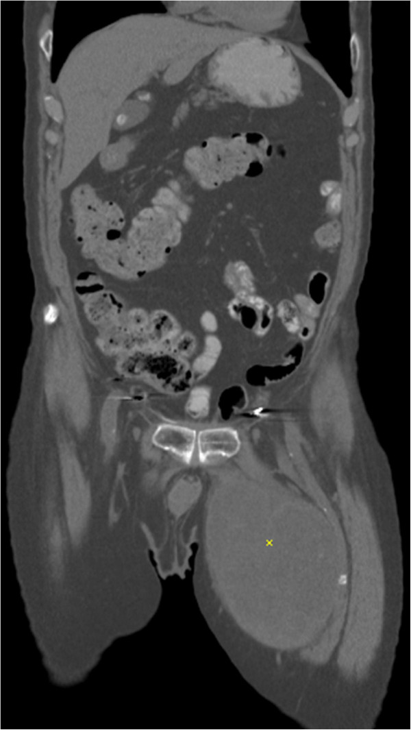
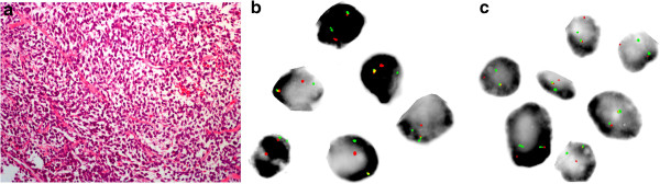
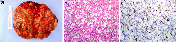
Results: We report a case of myxoid liposarcoma in a 91-year-old, the oldest known patient with this disease-entity. FISH analysis of the DDIT3 and FUS loci demonstrate the pathognomonic chromosomal alteration in the setting of predominantly round cell histology on biopsy, confirmed by RT-PCR.
Conclusion: Myxoid liposarcoma affects mostly young adults but can be seen in the elderly population. Molecular and cytogenetic assays are helpful auxiliaries to histology in the setting of unusual histology and clinical presentation.
Figures
Similar articles
-
DDIT3 gene break-apart as a molecular marker for diagnosis of myxoid liposarcoma--assay validation and clinical experience.Diagn Mol Pathol. 2011 Dec;20(4):218-24. doi: 10.1097/PDM.0b013e3182107eb9. Diagn Mol Pathol. 2011. PMID: 22089349
-
The utility of fluorescence in situ hybridization (FISH) in the diagnosis of myxoid soft tissue neoplasms.Am J Surg Pathol. 2008 Jan;32(1):8-13. doi: 10.1097/PAS.0b013e3181578d5a. Am J Surg Pathol. 2008. PMID: 18162764
-
A Patient with Myxoid/Round Cell Liposarcoma (MRCL) Involving the Well-known Translocation t(12;22): A Case Report with the Cytogenomic Landscape of this Rearrangement.J Assoc Genet Technol. 2022;48(2):68-75. J Assoc Genet Technol. 2022. PMID: 35660675
-
Myxoid liposarcoma with cartilaginous differentiation: A case study with fish analysis and review of the literature.Pathol Res Pract. 2013 Oct;209(10):666-9. doi: 10.1016/j.prp.2013.06.010. Epub 2013 Jul 12. Pathol Res Pract. 2013. PMID: 23920321 Review.
-
Vulvar Myxoid Liposarcoma and Well Differentiated Liposarcoma With Molecular Cytogenetic Confirmation: Case Reports With Review of Malignant Lipomatous Tumors of the Vulva.Int J Gynecol Pathol. 2015 Jul;34(4):390-5. doi: 10.1097/PGP.0000000000000170. Int J Gynecol Pathol. 2015. PMID: 25851712 Review.
Cited by
-
Dyspnea caused by a giant retroperitoneal liposarcoma: A case report.Oncol Lett. 2018 Aug;16(2):1539-1542. doi: 10.3892/ol.2018.8791. Epub 2018 May 24. Oncol Lett. 2018. PMID: 30008834 Free PMC article.
-
Oral liposarcoma in elderly: Case report and literature analysis.Medicine (Baltimore). 2020 Feb;99(6):e18985. doi: 10.1097/MD.0000000000018985. Medicine (Baltimore). 2020. PMID: 32028409 Free PMC article. Review.
-
Surgical removal of giant pelvic liposarcoma after preoperative transcatheter arterial embolization.Int Cancer Conf J. 2022 Aug 1;11(4):275-279. doi: 10.1007/s13691-022-00560-z. eCollection 2022 Oct. Int Cancer Conf J. 2022. PMID: 36186231 Free PMC article.
References
-
- Antonescu C, Ladanyi M. In: Pathology and genetics of tumours of soft tissue and bone. Fletcher CDM, Unni KK, Mertens F, editor. Lyon: IARC Press; 2002. Myxoid liposarcoma; pp. 40–43. [Kleihues P and Sobin LH (Series Editors): World Health Organization classification of tumors]
-
- Sreelantaiah C, Karakousis CP, Leong SPL, Sandberg AA. Cytogenetic findings in liposarcoma correlate with histopathologic subtypes. Cancer. 1991;69:2484–2495. - PubMed
Publication types
LinkOut - more resources
Full Text Sources
Other Literature Sources
Research Materials
