Comparative Study
. 2013 Nov 18;1(1):77.
doi: 10.1186/2051-5960-1-77.

The type of Aβ-related neuronal degeneration differs between amyloid precursor protein (APP23) and amyloid β-peptide (APP48) transgenic mice

Affiliations
Comparative Study

The type of Aβ-related neuronal degeneration differs between amyloid precursor protein (APP23) and amyloid β-peptide (APP48) transgenic mice

Ajeet Rijal Upadhaya et al. Acta Neuropathol Commun. .

Abstract

Background: The deposition of the amyloid β-peptide (Aβ) in the brain is one of the hallmarks of Alzheimer's disease (AD). It is not yet clear whether Aβ always leads to similar changes or whether it induces different features of neurodegeneration in relation to its intra- and/or extracellular localization or to its intracellular trafficking routes. To address this question, we have analyzed two transgenic mouse models: APP48 and APP23 mice. The APP48 mouse expresses Aβ1-42 with a signal sequence in neurons. These animals produce intracellular Aβ independent of amyloid precursor protein (APP) but do not develop extracellular Aβ plaques. The APP23 mouse overexpresses human APP with the Swedish mutation (KM670/671NL) in neurons and produces APP-derived extracellular Aβ plaques and intracellular Aβ aggregates.

Results: Tracing of commissural neurons in layer III of the frontocentral cortex with the DiI tracer revealed no morphological signs of dendritic degeneration in APP48 mice compared to littermate controls. In contrast, the dendritic tree of highly ramified commissural frontocentral neurons was altered in 15-month-old APP23 mice. The density of asymmetric synapses in the frontocentral cortex was reduced in 3- and 15-month-old APP23 but not in 3- and 18-month-old APP48 mice. Frontocentral neurons of 18-month-old APP48 mice showed an increased proportion of altered mitochondria in the soma compared to wild type and APP23 mice. Aβ was often seen in the membrane of neuronal mitochondria in APP48 mice at the ultrastructural level.

Conclusions: These results indicate that APP-independent intracellular Aβ accumulation in APP48 mice is not associated with dendritic and neuritic degeneration but with mitochondrial alterations whereas APP-derived extra- and intracellular Aβ pathology in APP23 mice is linked to dendrite degeneration and synapse loss independent of obvious mitochondrial alterations. Thus, Aβ aggregates in APP23 and APP48 mice induce neurodegeneration presumably by different mechanisms and APP-related production of Aβ may, thereby, play a role for the degeneration of neurites and synapses.


Figures
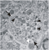
Figure 1
Figure 1
Identification of dystrophic neurites. Electron microscopy was used to identify dystrophic neurites (arrows) as previously described [19] and shown here in the frontocentral cortex of 15-month-old APP23 mice. Such neurites are characterized by neuritic swelling and contain vesicles with electron dense bodies (black arrowheads) probably representing autophagic vacuoles. Mitochondria in these neurites appear morphologically intact (M). Few multivesicular bodies are seen in these neurites as well (white arrowhead). The calibration bar corresponds to: 250 nm.
Figure 2
Figure 2
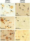
Aβ-pathology in 15-month-old APP23 (a, c, e) and 3-month-old-APP48 mice (b, d, f). a: The APP23 mouse showed a high number of extracellular Aβ plaques detectable with an antibody raised against Aβ17-24 (arrows). Intracellular Aβ was negligible. b: In the 3-month-old APP48 mouse no extracellular Aβ-pathology was apparent. These animals showed intraneuronal dendritic threads (arrowheads) and somatic granules (lucent arrow) as well as intramicroglial Aβ-grains (arrows) detectable with anti-Aβ17-24 as previously published [16]. c: Amyloid plaques in APP23 mice did also contain N-terminal truncated and pyroglutamate modified AβN3pE (arrows). d: AβN3pE was also found in even 3-month-old APP48 mice in some neuritic threads (arrowheads). e: phosphorylated Aβ (pAβ) was detected in amyloid plaques in 15-month-old APP23 mice. f: In 3-month-old APP48 mice only single threads showed labeling with anti-pAβ. Calibration bar in b corresponds to: a, b = 30 μm; c = 80 μm; e = 60 μm; d, f = 20 μm.
Figure 3
Figure 3
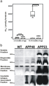
Biochemical analysis of Aβ in APP23 and APP48 mice. a: Total Aβ42 levels detected by ELISA in forebrain hemispheres of 2–3 and 15-18-month-old APP23 and APP48 mice. At 2–3 months APP23 mice exhibited low amounts of Aβ42 whereas APP48 mice displayed significantly more Aβ42 in the brain. At 15–18 months APP48 mice showed more Aβ than at 2–3 months of age but APP23 mice exhibited several times more Aβ in the forebrain. b: For demonstration of the types of Aβ aggregates in APP23 and APP48 mice brain homogenates of 9-11-month-old animals were analyzed by SDS-PAGE and western blotting after preparation of the soluble, dispersible, membrane-associated and insoluble (plaque-associated) fraction. Soluble Aβ as detected with antibodies raised against Aβ1-17 (6E10) was restricted to APP23 mice. Dispersible, membrane-associated, and insoluble (plaque-associated, formic acid soluble) Aβ aggregates were found in both transgenic mouse lines. The Aβ detected in the insoluble fraction of the forebrain homogenates of APP48 mice represents Aβ aggregates that require formic acid pretreatment before analysis similar to plaque-associated Aβ in APP23 mice. Since APP48 mice did not develop Aβ plaques this insoluble Aβ presumably represented intracellular fibrillar aggregates, such as dendritic threads. Wild type controls did not exhibit detectable amounts of Aβ in all four fractions. The original western blots are depicted in Additional file 3 (ELISA data from APP23 mice were previously published in a different context [18]). ***p < 0.001 Welch-test.
Figure 4
Figure 4
Dendritic degeneration in frontocentral commissural neurons of APP23 and APP48 mice. a: A type I neuron in an 18 month-old wild type animal exhibits a symmetric dendritic tree with prominent secondary and tertiary branches. b: In contrast, the dendritic tree of a representative type I commissural neuron (I) in a 15-month-old APP23 mouse is degenerated. Most basal dendrites were shrunken and had a reduced caliber (arrows). The degenerated dendrites showed some branches (arrows) that distinguished the degenerated type I neuron (I) from type II neurons without ramifications near the soma (II). c: Such a degeneration of the dendritic tree was not seen in APP48 mice. d-f: Numbers of DiI-traced type I, type II and type III commissural neurons in 15-18-month-old mice. d: APP23 mice at 15 months of age showed a decrease by more than 50% of the type I commissural neurons compared with 18-month-old wild type and APP48 mice. e: There was a significant reduction of type II commissural neurons in APP23 mice at 15 months of age compared with wild type littermates and APP48 mice at 18 months of age. f: Although APP23 mice had higher numbers of type III commissural neurons there was no significant difference from wild type littermates. g-h: No significant differences among the frequencies of DiI-traced type I, type II, and type III commissural neurons were observed at 3 months of age. ** p < 0.01 (Further statistical analysis: Additional file 2). Means and standard errors are depicted in d-i. (Quantitative data from APP23 mice and their respective wild type littermates were previously published in a different context [18]). Calibration bar in c corresponds to: a-c = 30 μm.
Figure 5
Figure 5
Frequencies of dystrophic neurites in wild type, APP23, and APP48 mice. The semiquantitatively assessed frequency of dystrophic neurites in the soma- and plaque-free frontocentral neuropil at the electron microscopic level was higher in 15-month-old APP23 mice than in wild type and APP48 mice of the same age group. In APP48 mice there was no increase in the frequency of dystrophic neurites in comparison to wild type mice. 3-month-old mice did not exhibit significant differences in the frequency of dystrophic neurites among the 3 genotypes nor were differences found in neuropil of the stratum radiatum and oriens of the CA1 region. (Data from APP23 mice and their respective wild type littermates were previously published in a different context [19]). *p < 0.05 (Further statistical analysis: Additional file 2). Means and standard errors are depicted.
Figure 6
Figure 6
Synapse densities in wild type, APP23 and APP48 mice. a: Loss of asymmetric synapses in the frontocentral cortex of 3- and 15-month-old APP23 mice in comparison to wild type mice. 18-month-old APP48 and wild type mice did not differ significantly in the density of asymmetric synapses. At 3 months of age APP48 mice had even more asymmetric synapses than wild type animals. In CA1 there were also slightly less asymmetric synapses in APP23 mice than in wild type controls and APP48 mice. However, these differences were not significant. b: There were no significant differences in the number of symmetric synapses in the frontocentral cortex and in CA1 in 3- and 15-18-month-old animals. c: APP23 and APP48 mice of both age groups exhibited reduced numbers of CA1 neurons compared to wild type mice whereby CA1 neuron loss was most pronounced in APP23 mice. d: The number of neurons in the frontocentral cortex did not vary significantly among 15-18-month-old wild type, APP23, and APP48 mice. Therefore, younger animals were not studied for the number of neurons in the frontocentral cortex. *p < 0.05, **p < 0.01, ***p < 0.001 (Further statistical analysis: Additional file 2). Means and standard errors are depicted. (Some data were previously published in a different context [16, 19]).
Figure 7
Figure 7
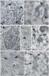
Electron microscopy and immunogold labeling of Aβ in APP23 and APP48 mice. a, b: Immunogold particles specifically labeled fibrillar Aβ (arrowheads) of a plaque in a 15-month-old APP23 mouse. At high magnification small amyloid fibrils were identified (arrowheads in b). They were located in the extracellular space. c, d: Dystrophic neurites (outlined structures) were associated with extracellular bundles of plaque-associated Aβ fibrils (arrowheads) in an 15-month-old APP23 mouse. Within the neurite, Aβ was localized in electron dense material near the surface as well as in the center of the neurite (arrows in c). d: A second dendrite without signs of dystrophy such as multilamellar bodies was also located near extracellular Aβ fibrils (outlined structure labeled with e). e: Higher magnification of this dendrite showed a dendrite cross section with an intact mitochondrium (m) and with condensed Aβ-positive material in the cytoplasm (arrows). Similar Aβ-positive material was found in the neighboring extracellular space (arrowhead). Both, intra- and extracellular Aβ aggregates did not exhibit fibrillar morphology. As such it is quite likely that these Aβ aggregates represent non-fibrillar oligomers and/or protofibrils occurring in the neighborhood of extracellular, plaque-associated Aβ fibrils. f: Fibrillar material (arrows) was observed in some dendrites of a 3-month-old APP48 mouse in an Epon-embedded, not immunostained section presumably representing the ultrastructural correlative for dendritic threads. g: Immunoelectron microscopy confirmed Aβ-positive material in fibrillar aggregates within dendritic threads labeled by gold particles (arrows) in APP48 mice. No Aβ was observed in the neighboring, non-altered mitochondrium (m). Calibration bar in g is valid for: a = 570 nm, b = 200 nm, c = 750 nm, d = 1000 nm, e = 275 nm, f, g = 350 nm.
Figure 8
Figure 8
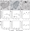
Electron microscopy of mitochondria in wild type, APP23, and APP48 mice. a-b: Electron microscopy showed predominantly non-altered mitochondria in nerve cell somata of wild type (WT) (a) and APP23 mice (b). c: More altered mitochondria in the nerve cell somata were observed in frontocentral neurons of 18-month-old APP48 mice. Mitochondrial alteration was characterized by a loss of mitochondrial christae although the double membrane architecture and at least single christa structures (arrow) were preserved. d-f: The percentage of altered mitochondria in the nerve cell somata of 18-month-old APP48 mice was higher than in 15-18-month-old wild type and APP23 mice (d). The volume density of altered mitochondria in the frontocentral nerve cell somata of 18-month-old APP48 mice was increased in comparison to 15-18-month-old wild type and APP23 mice (e). At 3 months of age such differences in the presence of altered mitochondria were not observed. The total mitochondrial volume density, i.e. altered and non-altered mitochondria together, did not differ among the investigated mouse lines in both age groups (f). g-i: In CA1 there were no obvious changes in the percentage and volume density of altered mitochondria in the nerve cell somata. *p < 0.05, **p < 0.01 (Further statistical analysis: Additional file 2). Means and standard errors are depicted in d-f. Calibration bar in c is valid for: a-c = 150 nm.
Figure 9
Figure 9
Immunoelectron microscopy of mitochondria in wild type, APP23, and APP48 mice. a: Immunoelectron microscopy revealed gold-labeled Aβ (arrows) within lipofuscin-like granules in the cytoplasm of frontocentral pyramidal neurons in 15-month-old APP23 mice. b. Most mitochondria in the somata of frontocentral pyramidal neurons in 15-month-old APP23 mice did not contain immunogold labeled Aβ-positive material (arrowheads). c: Only few neuronal mitochondria exhibited single gold particles in a 15-month-old APP23 mouse indicating anti-Aβ-positive material within these mitochondria (arrow). d: In a few mitochondria within frontocentral pyramidal nerve cell somata of 18-month-old APP48 mice we found immunogold labeled Aβ-positive material associated with the membranes of healthy-looking mitochondria (arrow). Neighboring non-altered mitochondria often did not contain Aβ (arrowheads). Aβ fibrils were not seen inside the mitochondria. e: Altered mitochondria within pyramidal neurons in the frontocentral cortex of a 18-month-old APP48 mouse also exhibited single gold particles in association with their membranes indicating the presence of Aβ (arrows) although Aβ fibrils were not observed inside the mitochondria. Due to the use of LR-white embedded tissue required for post-embedding immunoelectron microscopy (a,b) the tissue preservation was less good than in the Epon embedded sections used for the morphological analysis of the mitochondria (Figure 8 a-c). Calibration bar in e is valid for: a, b = 540 nm, c = 325 nm, d = 288 nm, e = 180 nm.
Figure 10
Figure 10
Schematic representation of the somatic and neuritic type of Aβ-related neurodegeneration in frontocentral neurons of APP23 and APP48 mice. In APP48 mice Aβ accumulates within neurons and in microglial cells as previously reported [16]. Extracellular Aβ is not detectable suggesting that APP-independently produced intracellular Aβ leads to functional impairment of neurons as indicated by motor deficits [16]. Mitochondrial alterations occur more frequently in the nerve cell somata of APP48 mice than in wild type and APP23 mice and are proposed to lead to apoptotic cell death as suggested previously [14] without preceding neuritic alteration. This type of somatic neurodegeneration in APP48 mice is different from that seen in APP23 mice, which contain less intracellular Aβ but significant amounts of extracellular Aβ aggregates including plaques. We propose that extra- and intracellular APP-derived Aβ causes synapse loss, dendrite degeneration and often plaque-associated, dystrophic neurites in APP23 mice indicative for a second neuritic type of neurodegeneration. Intracellular Aβ in APP23 mice may be produced within the nerve cell or may be taken up from the extracellular space [, –59].

Similar articles

Cited by

References

    1. Alzheimer A. Über eine eigenartige Erkrankung der Hirnrinde. Allg Zschr Psych. 1907;64:146–148.
    1. Masters CL, Simms G, Weinman NA, Multhaup G, McDonald BL, Beyreuther K. Amyloid plaque core protein in Alzheimer disease and Down syndrome. Proc Natl Acad Sci USA. 1985;82:4245–4249. doi: 10.1073/pnas.82.12.4245. - DOI - PMC - PubMed
    1. DeKosky ST, Scheff SW. Synapse loss in frontal cortex biopsies in Alzheimer’s disease: correlation with cognitive severity. Ann Neurol. 1990;27:457–464. doi: 10.1002/ana.410270502. - DOI - PubMed
    1. Masliah E, Terry RD, DeTeresa RM, Hansen LA. Immunohistochemical quantification of the synapse-related protein synaptophysin in Alzheimer disease. Neurosci Lett. 1989;103:234–239. doi: 10.1016/0304-3940(89)90582-X. - DOI - PubMed
    1. Terry RD, Peck A, DeTeresa R, Schechter R, Horoupian DS. Some morphometric aspects of the brain in senile dementia of the Alzheimer type. Ann Neurol. 1981;10:184–192. doi: 10.1002/ana.410100209. - DOI - PubMed

Publication types

MeSH terms