Comparative Study
. 2013 Jul 29:1:41.
doi: 10.1186/2051-5960-1-41.

Epilepsy: neuroinflammation, neurodegeneration, and APOE genotype

Affiliations
Comparative Study

Epilepsy: neuroinflammation, neurodegeneration, and APOE genotype

Orwa Aboud et al. Acta Neuropathol Commun. .

Abstract

Background: Precocious development of Alzheimer-type neuropathological changes in epilepsy patients, especially in APOE ϵ4,4 carriers is well known, but not the ways in which other APOE allelic combinations influence this outcome. Frozen and paraffin-embedded tissue samples resected from superior temporal lobes of 92 patients undergoing temporal lobectomies as a treatment for medication-resistant temporal lobe epilepsy were used in this study. To determine if epilepsy-related changes reflect those in another neurological condition, analogous tissue samples harvested from 10 autopsy-verified Alzheimer brains, and from 10 neurologically and neuropathologically normal control patients were analyzed using immunofluorescence histochemistry, western immunoblot, and real-time PCR to determine genotype effects on neuronal number and size, neuronal and glial expressions of amyloid β (Aβ) precursor protein (βAPP), Aβ, apolipoprotein E (ApoE), S100B, interleukin-1α and β, and α and β secretases; and on markers of neuronal stress, including DNA/RNA damage and caspase 3 expression.

Results: Allelic combinations of APOE influenced each epilepsy-related neuronal and glial response measured as well as neuropathological change. APOE ϵ3,3 conferred greatest neuronal resilience denoted as greatest production of the acute phase proteins and low neuronal stress as assessed by DNA/RNA damage and caspase-3 expression. Among patients having an APOE ϵ2 allele, none had Aβ plaques; their neuronal sizes, like those with APOE ϵ3,3 genotype were larger than those with other genotypes. APOE ϵ4,4 conferred the weakest neuronal resilience in epilepsy as well as in Alzheimer patients, but there were no APOE genotype-dependent differences in these parameters in neurologically normal patients.

Conclusions: Our findings provide evidence that the strength of the neuronal stress response is more related to patient APOE genotype than to either the etiology of the stress or to the age of the patient, suggesting that APOE genotyping may be a useful tool in treatment decisions.


Figures

Figure 1
Figure 1
The relationship between neuronal expression of ApoE and glial expression of IL-1α, relative to APOE genotype. IL-1α (green)-immunoreactive microglia clustered around individual ApoE-immunoreactive neurons (red) in patients with APOE ϵ2,3 (A), APOE ϵ4,4 (B), APOE ϵ3,4 (C), and APOE ϵ3,3 (D). A maximum of 9 microglia per neuron were counted in APOE ϵ3,3 patients (arrow in D); this was higher than the numbers associated with other APOE genotypes as shown in the percentage of neurons with adjacent IL-1α immunoreactive neurons (none to nine) relative to APOE genotype (E).
Figure 2
Figure 2
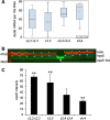
APOE genotype influences expression of the astrocyte-derived neuritogenic cytokine S100B. Wilcoxon distribution scores for S100B mRNA levels in patients with APOE ϵ4,4 (Group 4) is higher than that in other genotypes. p values: Group 4 vs 1, p = 0.004: Group 4 vs 2, p = 0.001; and Group 4 vs 3, p = 0.004 (A). S100B protein levels for all the patient samples (n = 92), quantified by western blot, showed that APOE ϵ3,3 patients (Group 2, n = 53) had higher levels of S100B than did other APOE genotypes (99.7 ± 5.17 vs 95.7 ± 3.55, 92.7 ± 6.21, 92.9 ± 2.87 arbitrary units; p = 0.001) for groups 1 (n = 13), 3 (n = 19), and 4 (n = 7), respectively (B). Illustration of S100B protein levels; one of eight western blots of different epilepsy samples (n = 92) with standard (Std) purified S100B positive control (middle sample S100B Std) the S100B mono band represents the S100B monomer (~11kD) (C).
Figure 3
Figure 3
Epilepsy-induced neuronal stress affects neuronal size in an APOE genotype-dependent fashion. Neurons in tissue from epilepsy patients were immunoreacted with a pan neuronal marker (green). The apparent large size of neurons in patients with APOE ϵ2,3 (A) and those in patients with APOE ϵ3,3 (B) relative to those in patients with APOE ϵ2,4 (C), APOE ϵ3,4 (D), and APOE ϵ4,4 genotype (E) was confirmed by measuring cross-sectional areas of neurons (μm2) in Groups 1,2,3,4, respectively (432 ± 40; 389 ± 29; 294 ± 20; vs 213 ± 17; p < 0.001) (F).
Figure 4
Figure 4
Cross sectional neuronal area in Alzheimer patients and in neurologically normal controls, relative to APOE genotype. Superior temporal gyrus tissue sections from Alzheimer patients (A-C) and neurologically normal patients (D-F) immunoreacted with Pan neuronal marker (green) in Alzheimer patients having APOE ϵ3,3 (A) appeared to be larger than those from Alzheimer patients with APOE ϵ4,4 (B). Cross-sectional area measurements of neurons (μm2) confirm this difference (286 ± 23, vs 227 ± 25; p = 0.009) (C). Cross-sectional area of neurons in control patients with APOE ϵ3,3 (D) was not different from that in control patients with APOE ϵ4,4 (E) (250 ± 16, vs 244 ± 28; p = 0.67) (F).
Figure 5
Figure 5
Neuronal stress as estimated by heat shock-related damage associated with expression of 8-OH guanosine. Numbers of neurons expressing RNA/DNA oxidative damage was similar in the different APOE genotype groups (A-C). However, the extent of damage per neuron, as assessed by Stress Marq (8-OH guanosine) fluorescence intensity, was greater in patients with APOE ϵ4,4 than APOE ϵ2,2 or ϵ3,3 genotypes (80 ± 3 vs 55 ± 6 and 55 ± 6 arbitrary units, p = 0.001) (D).
Figure 6
Figure 6
APOE genotype influences cell cycle-related DNA damage. Illustration of a western blot representative of 8 gels from patients (n = 92) with different APOE genotypes, showing actin bands (green) and pro-caspase 3 bands (red) (A). Wilcoxon distribution scores for pro-caspase 3 fluorescence intensity for all the 92 patients: APOE ϵ3,3 (Group 2) had lower levels of pro-caspase than other genotypes (B) with p values for comparisons with Groups 1,3,4 equal to < 0.002, < 0.001, and < 0.02, respectively.
Figure 7
Figure 7
ApoE transcription and translation relative to APOE genotype. Wilcoxon distribution scores for ApoE mRNA levels were higher in patients carrying at least one APOE ϵ4 allele (Group 3 and Group 4) than in those carrying other APOE genotypes (Groups 1 and 2); the difference was significant only between Group 2 and Group 3 (p < 0.01) (A). ApoE protein levels, illustrated in one of eight western blots of different epilepsy samples (n = 92) with actin (red) in the upper row and ApoE (green) in the lower row with the middle sample being recombinant ApoE protein used as a positive control (Std) (B). ApoE protein levels for all the patient samples (n = 92), quantified by western blot analysis, showed that APOE ϵ4,4 patients (Group 4) had lower levels of ApoE than did other APOE genotypes (18.2 ± 8.4 vs 61.8 ± 15.3, 55.4 ± 23.3, 26.9 ± 7.1 arbitrary units) for groups 1, 2, and 3, respectively, p < 0.01 (C).
Figure 8
Figure 8
Neuropathological changes in epilepsy relative to APOE genotype. Multiple IL-1α (green)/ApoE (red)-immunoreactive plaques were present in 13 of the 53 APOE ϵ3,3 patients (A) – the youngest of which was 17 yrs old – and in 3 of the 17 APOE ϵ3,4 patients (not shown here). One of the six APOE ϵ4,4 patients had Aβ plaques, a 10 year old male (B). The incidence of plaques in patients having each of the APOE allelic subtypes (C). Wilcoxon distribution scores for α-secretase mRNA measured relative to 18s mRNA showed that APOE ϵ4,4 patients (Group 4) had higher levels than did other APOE genotypes, with p values of 0.02, 0.01, and 0.01 for comparisons between group 4 and groups 1, 2, and 3, respectively (D).

References

    1. Chin JH. The global fund for epilepsy: a proposal. Neurology. 2013;1:754–755. doi: 10.1212/WNL.0b013e31828250c5. - DOI - PubMed
    1. Aboud O, Mrak RE, Boop F, Griffin ST. Apolipoprotein epsilon3 alleles are associated with indicators of neuronal resilience. BMC Med. 2012;1:35. doi: 10.1186/1741-7015-10-35. - DOI - PMC - PubMed
    1. Gouras GK, Relkin NR, Sweeney D, Munoz DG, Mackenzie IR, Gandy S. Increased apolipoprotein E epsilon 4 in epilepsy with senile plaques. Ann Neurol. 1997;1:402–404. doi: 10.1002/ana.410410317. - DOI - PubMed
    1. Mackenzie IR, Miller LA. Senile plaques in temporal lobe epilepsy. Acta Neuropathol (Berl) 1994;1:504–510. doi: 10.1007/BF00294177. - DOI - PubMed
    1. Annegers JF, Coan SP. The risks of epilepsy after traumatic brain injury. Seizure. 2000;1:453–457. doi: 10.1053/seiz.2000.0458. - DOI - PubMed

Publication types

MeSH terms

Substances