Comparative Study
. 2013 May 9;1(1):8.
doi: 10.1186/2051-5960-1-8.

Filamentous white matter prion protein deposition is a distinctive feature of multiple inherited prion diseases

Affiliations
Comparative Study

Filamentous white matter prion protein deposition is a distinctive feature of multiple inherited prion diseases

Lilla Reiniger et al. Acta Neuropathol Commun. .

Abstract

Background: Sporadic, inherited and acquired prion diseases show distinct histological patterns of abnormal prion protein (PrP) deposits. Many of the inherited prion diseases show striking histological patterns, which often associate with specific mutations. Most reports have focused on the pattern of PrP deposition in the cortical or cerebellar grey matter.

Results: We observed that the subcortical white matter in inherited prion diseases frequently contained filamentous depositions of abnormal PrP, and we have analysed by immunohistochemistry, immunofluorescence and electron microscopy 35 cases of inherited prion disease seen at the UK National Prion Clinic. We report here that filamentous PrP is abundantly deposited in myelinated fibres in inherited prion diseases, in particular in those with N-terminal mutations.

Conclusions: It is possible that the presence of filamentous PrP is related to the pathogenesis of inherited forms, which is different from those sporadic and acquired forms.


Figures
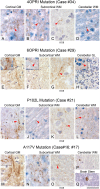
Figure 1
Figure 1
Threads of PrP positive deposits in inherited prion diseases with 4 and 6 octapeptide repeat insert mutation, P102L (Gerstmann Sträussler-Scheinker syndrome), and A117V mutation. AD, 4OPRI mutation with abundant threads, which form a dense crisscrossing network (corresponding to myelinated fibres) in the cortex (A) and forming multiple, thin parallel threads in the subcortical white matter, detectable with two antibodies, ICSM35 (B) and KG9 (C). Arrowheads in B and C point to accentuated threads. In the cerebellum (D), the PrP-containing filaments are thicker, shorter, and less abundant than in the cortex. The 6OPRI mutation (E-H) is characterised by abundant threads in the cortical grey matter I, subcortical white matter (F, ICSM35, G, KG9), and in the cerebellar granular layer (H). I-L, Both forebrain and cerebellum with PRNP P102L mutations show white matter threads of abnormal PrP in the subcortical (J, ICSM35 and K, KG9) or cerebellar (L) white matter. Arrows I-L show short threads of PrP deposition. Small myelinated fibres extending into the cerebral cortex also show threads of abnormal PrP (arrowhead in I) alongside with large amyloid plaques (not shown) and synaptic PrP. M-Q, A117V mutation (Case #16 M-P and Case #17, Q) with abundant cortical threads (M), corresponding to myelinated cortical fibres. The inset shows several parallel threads. Also the subcortical white matter is rich in PrP positive threads (N, ICSM35 and O, KG9). The cerebellum of the same case lacks filamentous inclusions, whilst small granular deposits are often observed (P). The brain stem of case #17 shows cross sections of myelinated fibres with occasional circular PrP-positive structures, corresponding to PrP containing myelin sheaths. Scale bar corresponds to 20 μm in P, 40 μm in the insets of E, F, H and M, to 80 μm in C, G, H, O and to 140 μm in all other panels.
Figure 2
Figure 2
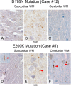
Patterns of PrP deposits in brains with PRNP D178N and E200K mutations. A-C, there are small dot like inclusions in the white matter of a case with D178N mutation (A, B, ICSM35 and KG9), no cerebellar threads (C). D-F, E200K mutation with sparse threads in the subcortical white matter (D, ICSM35 and E, KG9) and the cerebellar white matter (F). The inset in (D) shows a rare straight PrP positive filament and the arrowhead points at a short stub or dot of abnormal PrP. The cerebellar white matter (F) of the same case shows occasional thicker, straight filaments of abnormal PrP (arrowheads). Scale bar corresponds to 140 μm in (D), to 80 μm in B, E and to 40 μm in all other panels.
Figure 3
Figure 3
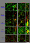
Co-localisation studies of abnormal PrP (Alexa 546, red) with Neurofilament (A, C, E, G, I) or with myelin basic protein (B, D, F, H, J), (green, Alexa 488) in the white matter of the frontal lobe. A, B, 4OPRI mutation shows the neurofilament signal being located beside abnormal PrP (A, A’), whereas there is an overlap between MBP and PrP signal (B, B’). C, D; 6OPRI with filamentous PrP being localised next to Neurofilament (C, C’) but directly congruent with MBP (D, D’). E, F there is little white matter PrP in this example of a P102L mutation, whilst a case with an A117V mutation (G, H) shows abundant white matter PrP, which shows PrP immunoreactivity next to neurofilaments, (G, G’) and co-localises with MBP, resulting in yellow signal (H, H’). The more C-terminal mutations, such as D178N show mostly granular PrP deposits and no strong filamentous PrP (I, J). Scale bar 16μm for A-J and 4 μm respectively for A’-J’.
Figure 4
Figure 4
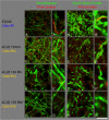
Co-localisation studies of abnormal PrP (Alexa 546, red) with Neurofilament (A, C, E, G) or with myelin basic protein (B, D, F, H), (green, Alexa 488) in the C-terminal E200K mutation and in sCJD control cases (frontal cortex). A, B, E200K mutations shows rare filamentous PrP, co-localising with the myelin sheath (B, B’) but not detectably with axonal neurofilament (A, A’). All sCJD cases (129VV, C, D; 129MV, E, F; 129MM, G, H) show variable amounts of granular PrP which does not co-localise with neurofilaments (C, E, G) or Myelin basic protein (D, F, H). Scale bar: 16 μm for A-H and 4 μm respectively for A’-H’.
Figure 5
Figure 5
Electron microscopy of two cases with abundant white matter deposition of PrP amyloid in the frontal cortex. A, B, Case #31 with 6OPRI mutation shows small granular deposits in the axoplasm of a longitudinal section of a myelinated axon, and more substantial fibrillar electron dense deposits in a myelin sheath, that has become divided by these inclusions, raising the possibility of amyloid. C, D; Case #17 with an A117V mutation shows a cross section of a myelinated fibre with the axon (Ax) being separated from the para-axonal inclusion that led to a splitting of the myelin (My) sheath. The red box indicates the region shown in D with a characteristic electron dense deposition, raising the possibility of amyloid. Scale Bars: 1 μm (A, C), 0.5 μm (B, D).
Figure 6
Figure 6
Schematic representation of the deposition of white matter filamentous PrP in relation to the mutation: the horizontal bar represents the PRNP open reading frame with the N terminus on the left and the C terminus on the right. OPRI and point mutations are indicated with red symbols. At each mutation, the cases with white matter threads (filaments) are indicated with stars; the number of stars indicates the abundance of deposits (corresponding to the score in Table 2). For cases where no filaments are seen, white matter dot-like deposits are indicated by filled circles and cases with no white matter deposits are symbolised by open circles. The blue number next to the symbols corresponds to the case numbers in Table 2, all figures and in the text). The position of the mutations on the schematic representation is not exactly to scale.
Figure 7
Figure 7
Regional distribution of filamentous PrP in a brain with A117V mutation. (Case #17): the intensity of the white matter filaments is represented in shades of red, the most intense representing a strong deposition (frontal, parietal and temporal brain) and there is a decreasing gradient towards the posterior lobes, the cerebellum and the brain stem, where fewer filaments were observed. The corresponding semi-quantitative evaluation is listed in Table 6.

Similar articles

Cited by

References

    1. Budka H, Aguzzi A, Brown P, Brucher JM, Bugiani O, Gullotta F, Haltia M, Hauw JJ, Ironside JW, Jellinger K, et al. Neuropathological diagnostic criteria for Creutzfeldt-Jakob disease (CJD) and other human spongiform encephalopathies (prion diseases) Brain Pathol. 1995;5:459–466. doi: 10.1111/j.1750-3639.1995.tb00625.x. - DOI - PubMed
    1. Liberski PP, Sikorska B, Hauw JJ, Kopp N, Streichenberger N, Giraud P, Boellaard J, Budka H, Kovacs GG, Ironside J, Brown P. Ultrastructural characteristics (or evaluation) of Creutzfeldt-Jakob disease and other human transmissible spongiform encephalopathies or prion diseases. Ultrastruct Pathol. 2010;34:351–361. doi: 10.3109/01913123.2010.491175. - DOI - PubMed
    1. Jansen C, Head MW, Rozemuller AJ, Ironside JW. Panencephalopathic Creutzfeldt-Jakob disease in the Netherlands and the UK: clinical and pathological characteristics of nine patients. Neuropathol Appl Neurobiol. 2009;35:272–282. doi: 10.1111/j.1365-2990.2008.01004a.x. - DOI - PubMed
    1. Mizutani T, Okumura A, Oda M, Shiraki H. Panencephalopathic type of Creutzfeldt-Jakob disease: primary involvement of the cerebral white matter. J Neurol Neurosurg Psychiatry. 1981;44:103–115. doi: 10.1136/jnnp.44.2.103. - DOI - PMC - PubMed
    1. Parchi P, Giese A, Capellari S, Brown P, Schulz-Schaeffer W, Windl O, Zerr I, Budka H, Kopp N, Piccardo P, et al. Classification of sporadic Creutzfeldt-Jakob disease based on molecular and phenotypic analysis of 300 subjects. Ann Neurol. 1999;46:224–233. doi: 10.1002/1531-8249(199908)46:2<224::AID-ANA12>3.0.CO;2-W. - DOI - PubMed

Publication types

MeSH terms

Supplementary concepts

LinkOut - more resources