. 2013 Aug 2:1:44.
doi: 10.1186/2051-5960-1-44.

Establishing quantitative real-time quaking-induced conversion (qRT-QuIC) for highly sensitive detection and quantification of PrPSc in prion-infected tissues

Affiliations

Establishing quantitative real-time quaking-induced conversion (qRT-QuIC) for highly sensitive detection and quantification of PrPSc in prion-infected tissues

Song Shi et al. Acta Neuropathol Commun. .

Abstract

Background: PrPSc, the only known constituent of prions, the infectious agents causing prion diseases, can be detected by real-time quaking-induced conversion (RT-QuIC). However, there is no efficient method to quantify the amount of PrPSc by RT-QuIC.

Results: Here we introduce quantitative RT-QuIC (qRT-QuIC) to quantify with high accuracy minute amounts of PrPSc in the brain and various peripheral tissues at levels far below detection by in vivo transmission. PrPSc is relatively resistant to treatment with proteinase K (PK). However, as there can also be a fraction of pathological PrP that is digested by PK, we use the term PrP27-30 to denote to the amount of PrPSc that can be detected by immunoblot after PK treatment. qRT-QuIC is based upon the quantitative correlation between the seeded amount of PrP27-30 and the lag time to the start of the conversion reaction detected by RT-QuIC. By seeding known amounts of PrP27-30 quantified by immunoblot into qRT-QuIC a standard calibration curve can be obtained. Based on this calibration curve, seeded undetermined amounts of PrP27-30 can be directly calculated. qRT-QuIC allowed to quantify PrP27-30 concentrations at extremely low levels as low as 10-15.5 g PrP27-30, which corresponds to 0.001 LD50 units obtained by in vivo i.c. transmission studies. We find that PrP27-30 concentration increases steadily in the brain after inoculation and can be detected at various time points during the incubation period in peripheral organs (spleen, heart, muscle, liver, kidney) in two experimental scrapie strains (RML, ME7) in the mouse.

Conclusions: We suggest that an automatic quantitative system to measure disease progression as well as prion contamination of organs, blood and food product is feasible. Moreover, the concept of qRT-QuIC should be applicable to measure other disease-associated proteins rich in β-pleated structures (amyloid) that bind ThT and that show seeded aggregation.


Figures

Figure 1
Figure 1
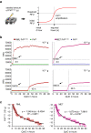
The basis of amplifying PrPSc with RT-QuIC. (a) Schematic illustration of RT-QuIC. PrPSc converts rPrPsen to rPrPres thereby increasing the total amount of β-sheeted PrP. This increase can be demonstrated by the increased value of ThT-fluorescence. Therefore, the sample containing PrPSc (prion) is distinguished from that without PrPSc (non-prion). RFU, relative fluorescence units. (b) However, it was not the amount of newly formed rPrPres but the time to reach the steep increase of amplification (ascendant curves) that was related to the seeded quantity of PK-treated PrPSc (PrP27-30). Different amounts of purified mouse RML scrapie-prion PrP27-30 and normal mouse PrPC were seeded into reactions to perform 80 hours of RT-QuIC. PrPC did not cause ascendant curves.
Figure 2
Figure 2
Directly detecting purified PrP27-30 with immunoblotting and ThT-binding fluorescence. (a) Purified mouse RML and ME7 scrapie-prion PrP27-30 was serially diluted and detected by immunoblotting. Aliquots were digested with 100 μg/ml Proteinase K (PK) before loading on the gel. Undigested PrPC is shown as a migration control. Signals were detected by 4H11 monoclonal antibody. Mr is shown on the right. (b) Fluorescence of purified RML and ME7 PrP27-30 and normal PrPC was measured after ThT-binding. The mean and s.e.m. are shown (n = 5).
Figure 3
Figure 3
Establishing the quantitative RT-QuIC (qRT-QuIC). (a) Schematic illustration of qRT-QuIC. The PrP27-30 propagating duration (hour) required to reach the threshold which was at least 3 times the starting fluorescence was set as the independent variable (x), the correlated seeded amount of PrP27-30 was the dependent variable (y). (b) Different amounts of purified PrP27-30 (10-10 to 10-16 g with serial 100.5-fold dilution) were seeded into RT-QuIC reactions using mouse rPrPsen as the substrate. The RT-QuIC process was followed from 0 to 90 h by showing the number of hours required to reach the threshold (indicated by black arrows and intersecting lines). Purified PrPC with identical amounts was seeded in independent RT-QuIC reactions as control. Seeded amounts of both PrP27-30 and PrPC are indicated on the top right; see also Additional file 1: Figures S2 and S3. (c) The results from five repeats of RT-QuIC seeded by PrP27-30 were provided to yield standard calibration curves and formulas for quantification. This relates QuIC time necessary to reach the threshold to the amount of seeded PrP27-30. The mean and s.e.m. are shown (n = 5).
Figure 4
Figure 4
Quantification of RML and ME7 PrP27-30 in brain at various days post infection (dpi). (a) 1 mg of brain from scrapie-infected mice at was analyzed by immunoblotting after digestion with 100 μg/ml PK. PrPC is shown as a migration control. Signals were detected by 4H11 monoclonal antibody. Mr is shown on the right. (b) Comparing PrP27-30 concentrations measured by quantitative RT-QuIC and quantitative immunoblot. The brains of RML-inoculated mice 90 dpi to 170 dpi and ME7-inoculated mice 90 to 150 dpi were chosen regarding the capacity of immunobloting to detect PrP27-30 from the time-point not earlier than 90 dpi. The significance (p value) was calculated with two-way ANOVA by using detecting methods and time-points as the variables. Shown data are mean and s.e.m (n = 5).
Figure 5
Figure 5
The seeding activity (expressed as PrP27-30 equvalents in g/g tissue) in various tissues from different time-points after inioculation were measured by qRT-QuIC by using the formulas shown in Figure3c. The PrP27-30 concentration corresponding to 1 LD50 units per gram of tissue is represented by a gray broken line. Shown are the mean and s.e.m. (n = 5).

Similar articles

Cited by

References

    1. Prusiner SB. Novel proteinaceous infectious particles cause scrapie. Science. 1982;1:136–144. doi: 10.1126/science.6801762. - DOI - PubMed
    1. Prusiner SB. Prions. Proc Natl Acad Sci USA. 1998;1:13363–13383. doi: 10.1073/pnas.95.23.13363. - DOI - PMC - PubMed
    1. Bieschke J, Weber P, Sarafoff N, Beekes M, Giese A, Kretzschmar H. Autocatalytic self-propagation of misfolded prion protein. Proc Natl Acad Sci USA. 2004;1:12207–12211. doi: 10.1073/pnas.0404650101. - DOI - PMC - PubMed
    1. Caughey B, Raymond GJ, Ernst D, Race RE. N-terminal truncation of the scrapie-associated form of PrP by lysosomal protease(s): implications regarding the site of conversion of PrP to the protease-resistant state. J Virol. 1997;1:6597–6603. - PMC - PubMed
    1. Pan KM, Baldwin M, Nguyen J, Gasset M, Serban A, Groth D, Mehlhorn I, Huang Z, Fletterick RJ, Cohen FE. Conversion of alpha-helices into beta-sheets features in the formation of the scrapie prion proteins. Proc Natl Acad Sci USA. 1993;1:10962–10966. doi: 10.1073/pnas.90.23.10962. - DOI - PMC - PubMed

Publication types