. 2013 Oct 2:1:63.
doi: 10.1186/2051-5960-1-63.

Human fetal inner ear involvement in congenital cytomegalovirus infection

Affiliations

Human fetal inner ear involvement in congenital cytomegalovirus infection

Liliana Gabrielli et al. Acta Neuropathol Commun. .

Abstract

Background: Congenital cytomegalovirus (CMV) infection is a leading cause of sensorineural hearing loss (SNHL). The mechanisms of pathogenesis of CMV-related SNHL are still unclear. The aim is to study congenital CMV-related damage in the fetal inner ear, in order to better understand the underlying pathophysiology behind CMV-SNHL.

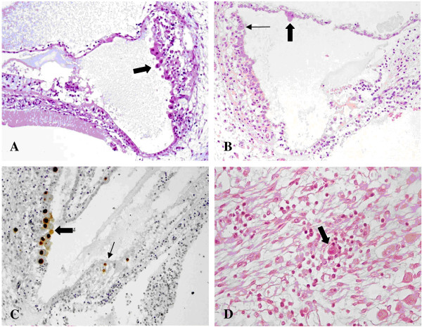
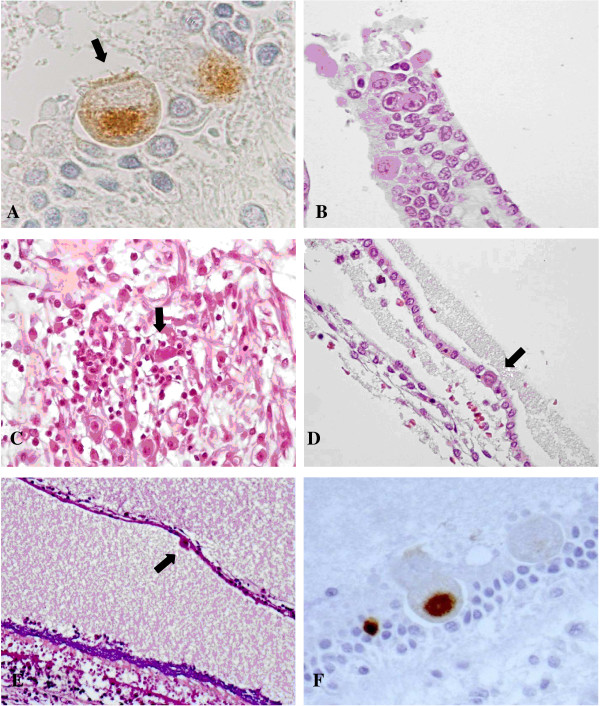
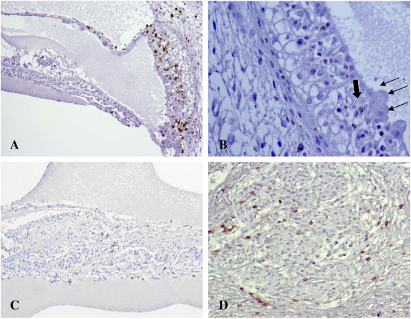
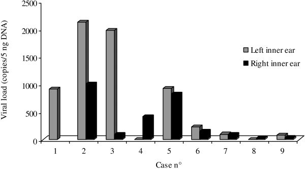
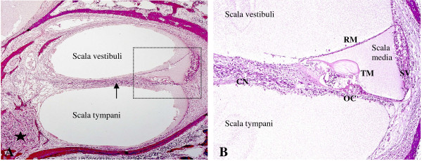
Results: We studied inner ears and brains of 20 human fetuses, all at 21 week gestational age, with a high viral load in the amniotic fluid, with and without ultrasound (US) brain abnormalities. We evaluated histological brain damage, inner ear infection, local inflammatory response and tissue viral load.Immunohistochemistry revealed that CMV was positive in 14/20 brains (70%) and in the inner ears of 9/20 fetuses (45%). In the cases with inner ear infection, the marginal cell layer of the stria vascularis was always infected, followed by infection in the Reissner's membrane. The highest tissue viral load was observed in the inner ear with infected Organ of Corti. Vestibular labyrinth showed CMV infection of sensory cells in the utricle and in the crista ampullaris.US cerebral anomalies were detected in 6 cases, and in all those cases, the inner ear was always involved. In the other 14 cases with normal brain scan, histological brain damage was present in 8 fetuses and 3 of them presented inner ear infection.

Conclusions: CMV-infection of the marginal cell layer of the stria vascularis may alter potassium and ion circulation, dissipating the endocochlear potential with consequent SNHL. Although abnormal cerebral US is highly predictive of brain and inner ear damage, normal US findings cannot exclude them either.


Figures

Figure 1
Figure 1
CMV infection in the cochlea. A) Numerous cytomegalic inclusions in the marginal layer of the stria vascularis (arrow). Haematoxylin and eosin (HE). B) Cytomegalic inclusions in the marginal layer of the stria vascularis (small arrow) and in the Reissner’s membrane (big arrow). HE. C) CMV immunohistochemistry showing strong nuclear CMV positivity in the marginal cell layer (big arrow) and in the Organ of Corti (small arrow). In the Organ of Corti, CMV-positive cells are most likely one ciliated cell (top) and one supporting cell (bottom). The other CMV-positive cell on the right is probably a supporting cell. D) Spiral ganglion: a cytomegalic neuron surrounded by lymphocytes. HE.
Figure 2
Figure 2
CMV infection in the vestibular apparatus. A) CMV immunohistochemistry showing CMV-positive cells within the utricle. The cell indicated by the arrow may likely be a sensory cell as suggested by the presence of cilia. B) Crista ampullaris: cytomegalic cells in the superficial epithelial layer. HE. C) Vestibular ganglion: a cluster of lymphocytes surrounding infected neurons (arrow). HE. D) One cytomegalic cell (arrow) in the epithelial layer of a semicircular canal. HE. E) Saccule: cytomegalic inclusion (arrow) in the membranous layer. On the bottom, there is the sensory macula with the otolith layer, strongly basophilic, and the hair cells underneath. HE. F) Saccule: cytomegalic cells within the macula. The positive cells may be a supporting cell (left) and a sensory cell (right). CMV immunohistochemistry.
Figure 3
Figure 3
Fetal inflammatory response in CMV-positive inner ears. A) CD8 immunohistochemistry showing numerous CD8-positive lymphocytes in the epithelial layers of the stria vascularis and within the Reissner’s membrane. Lymphocytic infiltrate was mainly found in proximity to cytomegalic cells. B) Granzyme B immunohistochemistry in the stria vascularis: most of the lymphocytes close to CMV infected cells (small arrows) were activated expressing Granzyme B (big arrow). C) CD8 immunohistochemistry showing CD8-positive lymphocytes along the cochlear nerve fibers. D) CD8 immunohistochemistry showing CD8-positive lymphocytes within the spiral ganglion.
Figure 4
Figure 4
Tissue viral load in the 15 CMV-positive inner ears.
Figure 5
Figure 5
Human fetal cochlea at 21 weeks gestation. A) Cochlear turn: the cochlea is divided in three tubular compartments: scala vestibuli, tympani and media (detail). The first two contain perilymph, a liquid with an ionic composition similar to extracellular fluids. The scala media contains endolymph, with a positive potential of 80 mV, the endocochlear potential, essential for stimulating the sensory cells of the Organ of Corti. The signals generated travel along the cochlear nerve fibers (arrow) to the spiral ganglion (star) and then through the auditory pathway. B) Scala media: the scala media contains the Organ of Corti (OC), composed of sensory cells innervated by the cochlear nerve (CN) and stimulated by the tectorial membrane (TM) according to endolymph waves. Endocochlear potential within the scala media is mainly maintained by the stria vascularis (SV) with the contribution of the Reissner’s membrane (RM).

Similar articles

Cited by

References

    1. Cheeran MC, Lokensgard JR, Schleiss MR. Neuropathogenesis of congenital cytomegalovirus infection: disease mechanisms and prospects for intervention. Clin Microbiol Rev. 2009;1:99–126. doi: 10.1128/CMR.00023-08. - DOI - PMC - PubMed
    1. Fowler KB, Boppana SB. Congenital cytomegalovirus (CMV) infection and hearing deficit. J Clin Virol. 2006;1:226–231. doi: 10.1016/j.jcv.2005.09.016. - DOI - PubMed
    1. Grosse SD, Ross DS, Dollard SC. Congenital cytomegalovirus (CMV) infection as a cause of permanent bilateral hearing loss: a quantitative assessment. J Clin Virol. 2008;1:57–62. doi: 10.1016/j.jcv.2007.09.004. - DOI - PubMed
    1. Rosenthal LS, Fowler KB, Boppana SB, Britt WJ, Pass RF, Schmid SD, Stagno S, Cannon MJ. Cytomegalovirus shedding and delayed sensorineural hearing loss: results from longitudinal follow-up of children with congenital infection. Pediatr Infect Dis J. 2009;1:515–520. doi: 10.1097/INF.0b013e318198c724. - DOI - PMC - PubMed
    1. Dahle AJ, Fowler KB, Wright JD, Boppana SB, Britt WJ, Pass RF. Longitudinal investigation of hearing disorders in children with congenital cytomegalovirus. J Am Acad Audiol. 2000;1:283–290. - PubMed

Publication types

MeSH terms