. 2013 Aug 9:1:48.
doi: 10.1186/2051-5960-1-48.

Immune complex formation impairs the elimination of solutes from the brain: implications for immunotherapy in Alzheimer's disease

Affiliations

Immune complex formation impairs the elimination of solutes from the brain: implications for immunotherapy in Alzheimer's disease

Roxana Octavia Carare et al. Acta Neuropathol Commun. .

Abstract

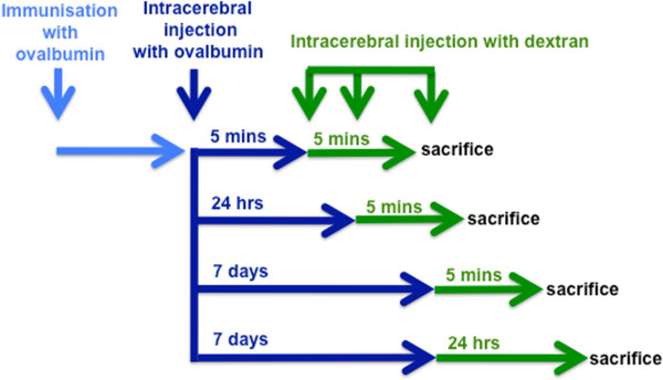
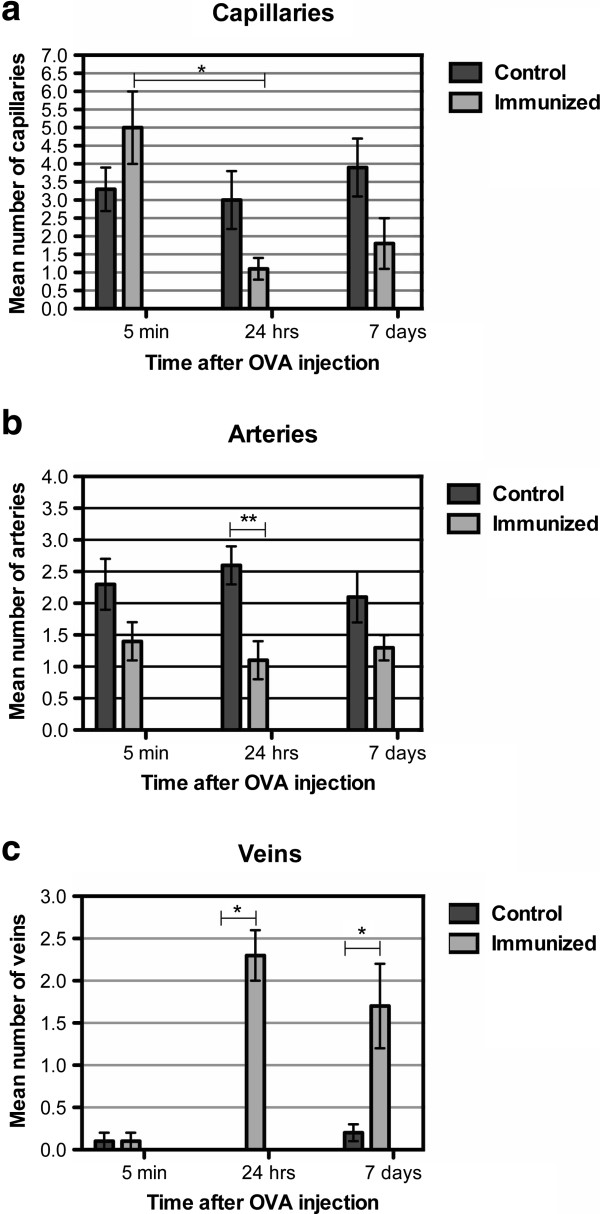
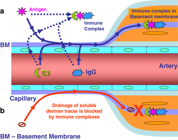
Background: Basement membranes in the walls of cerebral capillaries and arteries form a major lymphatic drainage pathway for fluid and solutes from the brain. Amyloid-β (Aβ) draining from the brain is deposited in such perivascular pathways as cerebral amyloid angiopathy (CAA) in Alzheimer's disease (AD). CAA increases in severity when Aβ is removed from the brain parenchyma by immunotherapy for AD. In this study we investigated the consequences of immune complexes in artery walls upon drainage of solutes similar to soluble Aβ. We tested the hypothesis that, following active immunization with ovalbumin, immune complexes form within the walls of cerebral arteries and impair the perivascular drainage of solutes from the brain. Mice were immunized against ovalbumin and then challenged by intracerebral microinjection of ovalbumin. Perivascular drainage of solutes was quantified following intracerebral microinjection of soluble fluorescent 3kDa dextran into the brain at different time intervals after intracerebral challenge with ovalbumin.

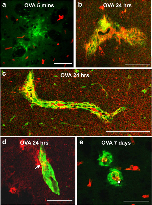
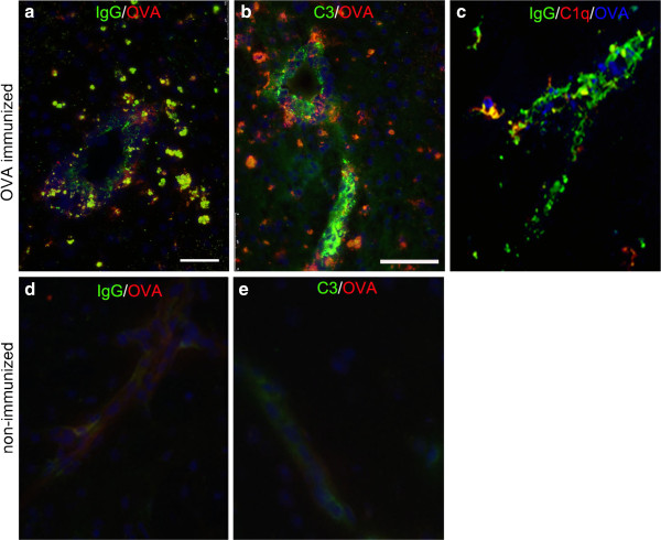
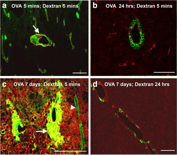
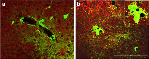
Results: Ovalbumin, IgG and complement C3 co-localized in basement membranes of artery walls 24 hrs after challenge with antigen; this was associated with significantly reduced drainage of dextran in immunized mice.

Conclusions: Perivascular drainage along artery walls returned to normal by 7 days. These results indicate that immune complexes form in association with basement membranes of cerebral arteries and interfere transiently with perivascular drainage of solutes from the brain. Immune complexes formed during immunotherapy for AD may similarly impair perivascular drainage of soluble Aβ and increase severity of CAA.


Figures

Figure 1
Figure 1
Schematic demonstrating the temporal sequence of immunization and injection experiments. Wild type BALB/c mice were immunized with OVA and then injected with OVA in the striatum at different time points. A soluble fixable fluorescent dextran was then injected intracerebrally at 5 mins or 24 h or 7 days post-immunization. Mice were examined at 5 minutes after injections of dextran.
Figure 2
Figure 2
Immune complexes in basement membranes in the walls of cerebral capillaries and arteries. Active immunization with OVA was followed by intracerebral challenge with OVA and left in situ for (a) 5 mins, (b-d) 24 hours and (e) 7 days. (a) At 5 mins, complement C3 (green) is diffusely in the vicinity of blood vessels stained for laminin (red); there is no co-localization of complement and laminin. (b) At 24 hours, IgG (red) is seen distributed diffusely in the perivascular brain parenchyma and is co-localized (yellow) with complement C3 (green) in brain parenchyma. (c) At 24 hours after injection of antigen, a longitudinal section of an artery in the striatum shows co-localization (yellow) of complement (green) with laminin (red) in the basement membranes surrounding smooth muscle cells in the tunica media. (d) IgG (red) is present in the brain parenchyma and co-localizes (yellow) with laminin (green) in a blood vessel wall at 24 hours. (e) At 7 days, complement C3 (green) is distributed circumferentially around blood vessels (red=laminin). Some co-localization of complement C3 and laminin (yellow) is still seen in the blood vessel walls. Confocal images: co-localization appears as a yellow colour. Scale bars = 60 μm.
Figure 3
Figure 3
Immune complexes. Immune complex formation was analyzed at 24 h after intracerebral OVA injection and stained for: a and d) IgG(green) and OVA (red), b and e) C3 (green) and OVA (red) and c) IgG (green), C1q (red) and OVA (blue). Co-localization is shown in yellow. Representative of n=3. Scale bar =75 am.
Figure 4
Figure 4
Perivascular drainage of dextran. (a) Following active immunization with OVA, at 5 minutes after the intracerebral injection of OVA and 5 minutes after the injection of dextran (green), the distribution of dextran is diffuse in the extracellular spaces and within the walls of capillaries and arteries. The arrow points to an artery with smooth muscle staining (red). (b) Following active immunization with OVA, at 24 hours after the intracerebral injection of OVA, the dextran injected intracerebrally (green) is present within 5 minutes of its injection as cuffs around vascular laminin (red). (c) In mice actively immunized with OVA, at 7 days after the intracerebral injection of OVA, dextran (green) is in the walls of blood vessels (arrowhead) and present within cuffs around large blood vessels (arrow). (d) In mice actively immunized with OVA, at 7 days after the intracerebral injection of OVA and 24 h after the intracerebral injection of dextran (green), the dextran is present in cells within a perivascular location (red-laminin). Confocal images: co-localization appears as a yellow colour. Scale bars: a, b, d = 100 μm; c = 120 μm.
Figure 5
Figure 5
Quantification of the number of blood vessels labeled with fluorescent dextran following the formation of immune complexes. Control (non-immunized) and OVA-immunized mice where OVA was left intracerebrally for 5 minutes, 24 h, 7 days, followed by the injection of fluorescent dextran and animals sacrificed within 5 mins. (a) The number of capillaries in control and OVA immunized mice, following intracerebral injection of OVA at 5 minutes, 24 h, or 7 days. The number of capillaries labeled with dextran is significantly lower in immunized animals, analyzed 24 h after OVA injection, compared to analysis at 5 minutes. (b) The number of arteries in control and OVA immunized mice, following intracerebral injection of OVA at 5 minutes, 24 h, or 7 days. The number of arteries labeled with dextran is significantly lower in OVA immunized mice where immune complexes were left to form for 24 hours, compared to the control non-immunized group. (c) There is a significant difference between the number of veins with dextran in a perivascular location in the OVA immunized mice analyzed at 24 h or 7 days, compared to control non-immunized mice. *p≤0.001; **p<0.005.
Figure 6
Figure 6
Active immunization with OVA followed by intracerebral challenge with OVA that was then left in situ for 7 days. Immune complex formation induces expression of F4/80 on perivascular macrophages and microglia and changes the pattern for the diffusion and elimination of dextran. (a) In non-immunized control mice, dextran (green) outlines the contour of a large blood vessel. Immunofluorescence for F4/80 reveals no staining. (b) 7 days following immune complex formation, F4/80 staining (red) is seen in the parenchyma and adjacent to blood vessels. Dextran (green) forms a large cuff around a blood vessel and is closely associated with the staining for F4/80. The yellow staining within the dextran cuff (inset) indicates co-localization with F4/80. Scale bars = 100 μm.
Figure 7
Figure 7
Diagram to illustrate the possible mechanism by which immune complexes disrupt the perivascular drainage of solutes. In (a), soluble antigen in the extracellular space interacts with IgG extravasated from the circulation resulting in immune complex formation and fixation of complement C3. The immune complexes formed block the arterial basement membranes that represent the perivascular drainage pathway. In (b), the drainage of a soluble tracer (dextran) is blocked by the presence of immune complexes in the arterial basement membranes. Alternatively, dextran is taken up by activated macrophages and microglia surrounding the blood vessels. We previously showed that macrophages/microglia are activated by FcR ligation following immune complex formation, which initiates an inflammatory in response surrounding the blood vessels (veins). The activated macrophages take up dextran by phagocytosis, thereby indirectly impeding elimination of solutes from the brain.

References

    1. Cserr HF, Cooper DN, Suri PK, Patlak CS. Efflux of radiolabeled polyethylene glycols and albumin from rat brain. AmJ Physiol. 1981;1(4):F319–F28. - PubMed
    1. Carare RO, Bernardes-Silva M, Newman TA, Page AM, Nicoll JA, Perry VH. et al.Solutes, but not cells, drain from the brain parenchyma along basement membranes of capillaries and arteries: significance for cerebral amyloid angiopathy and neuroimmunology. NeuropatholApplNeurobiol. 2008;1(2):131–44. - PubMed
    1. Weller RO, Boche D, Nicoll JA. Microvasculature changes and cerebral amyloid angiopathy in Alzheimer's disease and their potential impact on therapy. Acta neuropathologica. 2009;1(1):87–102. doi: 10.1007/s00401-009-0498-z. - DOI - PubMed
    1. Attems J. Sporadic cerebral amyloid angiopathy: pathology, clinical implications, and possible pathomechanisms. Acta Neuropathol(Berl) 2005;1(4):345–59. doi: 10.1007/s00401-005-1074-9. - DOI - PubMed
    1. Attems J, Jellinger K, Thal DR, Van Nostrand W. Review: sporadic cerebral amyloid angiopathy. Neuropathology and applied neurobiology. 2011;1(1):75–93. doi: 10.1111/j.1365-2990.2010.01137.x. Epub 2010/10/16. - DOI - PubMed

Publication types