Comparative Study
. 2013 Jul 1:1:29.
doi: 10.1186/2051-5960-1-29.

Comparative utility of LC3, p62 and TDP-43 immunohistochemistry in differentiation of inclusion body myositis from polymyositis and related inflammatory myopathies

Affiliations
Comparative Study

Comparative utility of LC3, p62 and TDP-43 immunohistochemistry in differentiation of inclusion body myositis from polymyositis and related inflammatory myopathies

Annie Hiniker et al. Acta Neuropathol Commun. .

Abstract

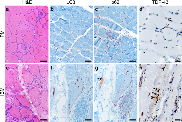
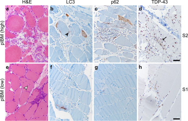
Background: Inclusion body myositis (IBM) is a slowly progressive inflammatory myopathy of the elderly that does not show significant clinical improvement in response to steroid therapy. Distinguishing IBM from polymyositis (PM) is clinically important since PM is steroid-responsive; however, the two conditions can show substantial histologic overlap.

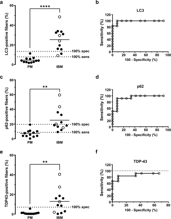
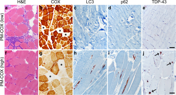
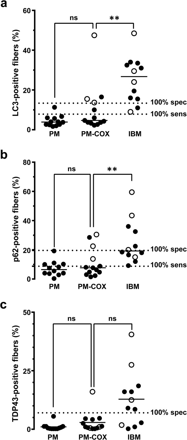
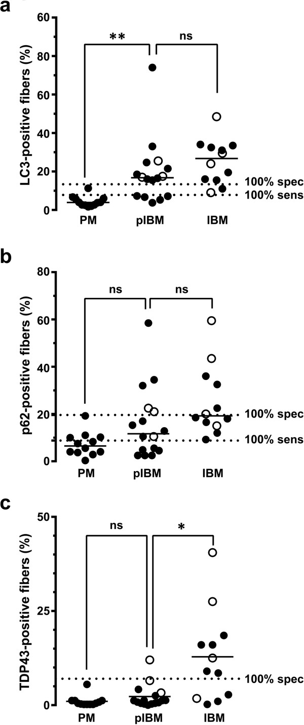
Results: We performed quantitative immunohistochemistry for (1) autophagic markers LC3 and p62 and (2) protein aggregation marker TDP-43 in 53 subjects with pathologically diagnosed PM, IBM, and two intermediate T cell-mediated inflammatory myopathies (polymyositis with COX-negative fibers and possible IBM). The percentage of stained fibers was significantly higher in IBM than PM for all three immunostains, but the markers varied in sensitivity and specificity. In particular, both LC3 and p62 were sensitive markers of IBM, but the tradeoff between sensitivity and specificity was smaller (and diagnostic utility thus greater) for LC3 than for p62. In contrast, TDP-43 immunopositivity was highly specific for IBM, but the sensitivity of this test was low, with definitive staining present in just 67% of IBM cases.

Conclusions: To differentiate IBM from PM, we thus recommend using a panel of LC3 and TDP-43 antibodies: the finding of <14% LC3-positive fibers helps exclude IBM, while >7% of TDP-43-positive fibers strongly supports a diagnosis of IBM. These data provide support for the hypothesis that disruption of autophagy and protein aggregation contribute to IBM pathogenesis.


Figures

Figure 1
Figure 1
PM and IBM staining patterns. A representative case of PM (a-d; subject #10) shows endomysial lymphocytic inflammation and muscle fiber invasion but no chronic myopathic features (a; H&E stain of the frozen material). There is no significant sarcoplasmic staining with LC3 (b), p62 (c), or TDP-43 (d), but TDP-43 stain highlights a subset of myofiber and inflammatory cell nuclei (internal positive control), while p62 faintly stains a subset of lymphocytes. A representative case of IBM (e-h, subject #22) shows endomysial inflammation accompanied by moderate to severe endomysial fibrosis, muscle fiber size variation, and RVs (white arrowhead) (e; H&E, frozen material). Staining for LC3 (f) and p62 (g) highlights RV rims; p62 also labels RV-associated protein aggregates (arrow) and scattered lymphocytes. TDP-43 immunostain (h) labels sarcoplasmic threads/skeins (black arrowheads), large protein aggregates (arrows), and coarse background puncta. Scale bars, 50 μM for a-c and e-g; 20 μM for d and h.
Figure 2
Figure 2
Quantification of LC3, p62, and TDP-43 positive fibers in the PM and IBM groups. The percentage of LC3- (a), p62- (c), and TDP-43-positive fibers (e) was significantly higher in the IBM group than the PM group. Each subject is represented with a symbol; the open symbols indicate subjects with known IBM clinical presentation. The unbroken lines designate group means, while dotted lines mark 100% sensitivity and 100% specificity cutoffs for each marker derived from ROC analysis. ****, p < 0.0001; **, p < 0.01. ROC analysis (b, d, and f) shows that quantitative immunohistochemistry for each of the three markers successfully differentiates IBM from PM subjects (p ≤ 0.001), although with varying tradeoffs between specificity and sensitivity. Of the three markers examined, only TDP-43 (f) failed to reach 100% sensitivity threshold, indicated by the dotted line in (f).
Figure 3
Figure 3
PM-COX staining patterns. PM-COX cases were histologically similar to classic PM, with endomysial inflammation, fiber invasion, and lack of well-developed chronic myopathic features (a and f; H&E, frozen material), but with ≥ 1% COX-negative fibers (b and g; COX stain, frozen material; COX-negative fibers are marked by asterisks). The majority of PM-COX samples (designated PM-COX low) showed no significant sarcoplasmic labeling for LC3 (c), p62 (d), or TDP-43 (e). In a subset of samples (designated PM-COX high), LC3 labeled RV rims (h), p62 labeled RV rims and small protein aggregates (i), while TDP-43 labeled sarcoplasmic skeins and large protein aggregates (arrows; j). a-e, subject #37, f-j, subject #33; scale bar, 50 μM.
Figure 4
Figure 4
Quantification of LC3, p62, and TDP-43 positive fibers in the PM-COX group. The percentage of LC3- (a) and p62-positive fibers (b) was significantly lower in the PM-COX group than in the IBM group, but similar to the PM group. With TDP-43 (c), there was no statistically significant difference between the PM-COX group and either the PM or IBM group. Each subject is represented with a symbol; the open symbols indicate subjects with known IBM clinical presentation. The unbroken lines designate group medians, while the dashed lines mark 100% sensitivity and 100% specificity cutoffs for each marker (derived from the ROC analysis shown in Figure 2). **, p < 0.01.
Figure 5
Figure 5
pIBM staining patterns. pIBM cases were histologically similar to classic IBM, with endomysial inflammation, fiber invasion, and moderate-severe chronic myopathic features, but without classic RVs (a and f; H&E, frozen material); “rimmed cracks” (white arrowhead in e) were present in a subset of specimens. The majority of pIBM samples (designated pIBM high) showed well-developed labeling for LC3 (b) and p62 (c); TDP-43 immunopositivity was less commonly observed (d; arrow marks a single TDP-43 positive fiber). In the example shown (subject #52), many fibers showed dense coarse puncta and rare RV-like structures (black arrowhead in b). In a smaller subset of samples (designated pIBM low), little or no labeling was seen with all three markers (f-h). a-d, subject #52, e-h, subject #51; scale bar, 50 μM.
Figure 6
Figure 6
Quantification of LC3, p62, and TDP-43 positive fibers in the pIBM group. The percentage of LC3-positive fibers (a) was significantly higher in the pIBM group than in the PM group, but similar to the IBM group. With p62 (b), there was no statistically significant difference between the pIBM group and either the PM or IBM group. The percentage of TDP-43-positive fibers (c) was significantly lower in the pIBM group than in the IBM group, but similar to the PM group. Each subject is represented with a symbol; the open symbols indicate subjects with known IBM clinical presentation. The unbroken lines designate group medians, while the dotted lines mark 100% sensitivity and 100% specificity cutoffs for each marker (derived from the ROC analysis shown in Figure 2). **, p < 0.01; *, p < 0.05.

References

    1. Dalakas MC. Sporadic inclusion body myositis–diagnosis, pathogenesis and therapeutic strategies. Nat Clin Pract Neurol. 2006;1:437–447. doi: 10.1038/ncpneuro0261. - DOI - PubMed
    1. Dalakas MC. Review: An update on inflammatory and autoimmune myopathies. Neuropathol Appl Neurobiol. 2011;1:226–242. doi: 10.1111/j.1365-2990.2010.01153.x. - DOI - PubMed
    1. Greenberg SA. Inclusion body myositis. Curr Opin Rheumatol. 2011;1:574–578. doi: 10.1097/BOR.0b013e32834b53cc. - DOI - PubMed
    1. Askanas V, Engel WK, Nogalska A. Pathogenic considerations in sporadic inclusion-body myositis, a degenerative muscle disease associated with aging and abnormalities of myoproteostasis. J Neuropathol Exp Neurol. 2012;1:680–693. doi: 10.1097/NEN.0b013e31826183c8. - DOI - PubMed
    1. Garlepp MJ, Mastaglia FL. Inclusion body myositis: new insights into pathogenesis. Curr Opin Rheumatol. 2008;1:662–668. doi: 10.1097/BOR.0b013e328313644c. - DOI - PubMed

Publication types

MeSH terms