Comparative Study
. 2013 Jul 6:1:31.
doi: 10.1186/2051-5960-1-31.

The unfolded protein response is activated in disease-affected brain regions in progressive supranuclear palsy and Alzheimer's disease

Collaborators, Affiliations
Comparative Study

The unfolded protein response is activated in disease-affected brain regions in progressive supranuclear palsy and Alzheimer's disease

Lauren D Stutzbach et al. Acta Neuropathol Commun. .

Abstract

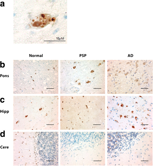
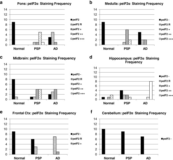
Background: Progressive supranuclear palsy (PSP) is a neurodegenerative disorder pathologically characterized by intracellular tangles of hyperphosphorylated tau protein distributed throughout the neocortex, basal ganglia, and brainstem. A genome-wide association study identified EIF2AK3 as a risk factor for PSP. EIF2AK3 encodes PERK, part of the endoplasmic reticulum's (ER) unfolded protein response (UPR). PERK is an ER membrane protein that senses unfolded protein accumulation within the ER lumen. Recently, several groups noted UPR activation in Alzheimer's disease (AD), Parkinson's disease (PD), amyotrophic lateral sclerosis, multiple system atrophy, and in the hippocampus and substantia nigra of PSP subjects. Here, we evaluate UPR PERK activation in the pons, medulla, midbrain, hippocampus, frontal cortex and cerebellum in subjects with PSP, AD, and in normal controls.

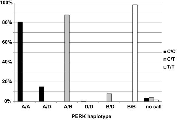
Results: We found UPR activation primarily in disease-affected brain regions in both disorders. In PSP, the UPR was primarily activated in the pons and medulla and to a much lesser extent in the hippocampus. In AD, the UPR was extensively activated in the hippocampus. We also observed UPR activation in the hippocampus of some elderly normal controls, severity of which positively correlated with both age and tau pathology but not with Aβ plaque burden. Finally, we evaluated EIF2AK3 coding variants that influence PERK activation. We show that a haplotype associated with increased PERK activation is genetically associated with increased PSP risk.

Conclusions: The UPR is activated in disease affected regions in PSP and the genetic evidence shows that this activation increases risk for PSP and is not a protective response.


Figures

Figure 1
Figure 1
pPERK is activated in PSP, AD, and normal brain. a. Example of a cell with pPERK immunoreactive puncta in the pons of a PSP case. b-d. Representative fields showing pPERK staining of pons, hippocampus, and cerebellum in normal, PSP, and AD cases. Scale bars are 50 μm unless otherwise indicated.
Figure 2
Figure 2
peIF2α is activated in PSP, AD, and normal brain. a. Example of a cell with peIF2α puncta in the pons of a PSP case. b-d. Representative fields from peIF2α staining of pons, hippocampus, and cerebellum in normal, PSP and AD cases. Scale bars are 50 μm unless otherwise indicated.
Figure 3
Figure 3
Frequency of pPERK staining scores in PSP, AD, and normal brain. Distribution of pPERK staining scores. +++ = widespread activation, ++ = moderate activation, + = diffuse activation, R = rare activation, - = no activation. Y-axis indicates number of cases with a particular pPERK staining score. All P-values obtained using Fisher exact test. a-c. PSP cases had the strongest pPERK staining in the pons (PSP vs. Normal: p = 3.8E-9) and the medulla (PSP vs. Normal: p = 6.1E-7), as well as moderate staining in the midbrain (PSP vs. Normal: p = 6.0E-6) which was affected in all PSP cases. d-e. AD cases had the strongest pPERK staining in the hippocampus (AD vs. Normal: p = 0.0006) and moderate staining in the frontal cortex (AD vs. Normal: p = 4.1E-5) both of which were affected in AD. f. No cases had significant pPERK staining in the cerebellum.
Figure 4
Figure 4
Frequency of peIF2α staining scores in PSP, AD, and normal brain. Distribution of peIF2α staining scores. +++ = widespread activation, ++ = moderate activation, + = diffuse activation, R = rare activation, - = no activation. Y-axis indicates number of cases with a particular peIF2α staining score. All P-values obtained using Fisher exact test. a-c. PSP cases had the strongest peIF2α staining in the pons (PSP vs. Normal: p = 4.1E-5) and the medulla (PSP vs. Normal: p = 6.0E-6), as well as moderate staining in the midbrain (PSP vs. Normal: p = 0.00041) which was affected in all PSP cases. d-e. AD cases had the strongest peIF2α staining in the hippocampus (AD vs. Normal: p = 0.0042) and moderate staining in the frontal cortex (AD vs. Normal: p = 4.1E-5) both of which were affected in AD. f. No cases had significant peIF2α staining in the cerebellum.
Figure 5
Figure 5
Hyperphosphorylated tau and pPERK partially co-localize in PSP pons and AD hippocampus. a. Example of a neuron co-stained for htau (red) and pPERK (green). Tau staining is widespread and diffuse. pPERK staining is punctate and localized to the soma and proximal neurites. b. pPERK staining occurred mostly in cells with diffuse, non-fibrillar htau staining. Cells with dense, fibrillar htau staining did not stain for activated PERK (*). c. In PSP pons, most pPERK positive cells also stained for htau (72%), whereas fewer than half of htau stained cells (43%) also stained positive for pPERK. d. htau and pPERK staining overlapped very little in AD hippocampus (14% and 20%). Scale bars are 50 μm unless otherwise indicated.
Figure 6
Figure 6
Severity of PERK activation in normal hippocampus correlates with age and tau pathology. a. Plot of pPERK staining score (X-axis) versus subject age at death (Y-axis). Each diamond represents one normal subject. Some individuals both young and old were negative for pPERK staining. Of those that stained positive for pPERK (including those showing rare through +++ levels of immunoreactivity), older individuals tended to have more severe pPERK staining scores. b. Frequency table plotting htau score against pPERK staining score in normal hippocampus. Htau score and pPERK score were positively correlated (Spearman R: .7523; p = 0.0002). The higher the htau score of an individual hippocampus, the higher the pPERK staining score tended to be. Htau scores were obtained from the CNDR Integrated Neurodegenerative Disease Database [33] using antibody PHF-1.
Figure 7
Figure 7
Comparison of PERK haplotype with GWAS risk allele. A GWAS for PSP identified a risk locus on chromosome 2 (rs7571971). The common, low risk allele at this locus is cytosine (C) and the PSP risk allele is thymine (T) [10]. Among individuals homozygous for C at this locus, all harbor PERK haplotype A or D in some combination. Individuals heterozygous (C/T) at this locus were heterozygous for haplotypes A, B, and/or D. Individuals homozygous for T at the GWAS risk locus were always homozygous for PERK haplotype B. This demonstrates that one of the two amino acid changes conferred by the B PERK haplotype that are not shared by the D haplotype may be responsible for the PSP risk evident on Chr. 2.

Similar articles

Cited by

References

    1. Litvan I, Agid Y, Calne D, Campbell G, Dubois B, Duvoisin RC, Goetz CG, Golbe LI, Grafman J, Growdon JH. et al.Clinical research criteria for the diagnosis of progressive supranuclear palsy (Steele-Richardson-Olszewski syndrome): report of the NINDS-SPSP international workshop. Neurology. 1996;1:1–9. doi: 10.1212/WNL.47.1.1. - DOI - PubMed
    1. Steele JC, Richardson JC, Olszewski J. Progressive supranuclear palsy. A heterogeneous degeneration involving the brain stem, basal ganglia and cerebellum with vertical gaze and pseudobulbar palsy, nuchal dystonia and dementia. Arch Neurol. 1964;1:333–359. doi: 10.1001/archneur.1964.00460160003001. - DOI - PubMed
    1. Dickson DW, Rademakers R, Hutton ML. Progressive supranuclear palsy: pathology and genetics. Brain Pathol. 2007;1:74–82. doi: 10.1111/j.1750-3639.2007.00054.x. - DOI - PMC - PubMed
    1. Ballatore C, Lee VMY, Trojanowski JQ. Tau-mediated neurodegeneration in Alzheimer’s disease and related disorders. Nat Rev Neurosci. 2007;1:663–672. - PubMed
    1. Stanford PM, Halliday GM, Brooks WS, Kwok JB, Storey CE, Creasey H, Morris JG, Fulham MJ, Schofield PR. Progressive supranuclear palsy pathology caused by a novel silent mutation in exon 10 of the tau gene: expansion of the disease phenotype caused by tau gene mutations. Brain. 2000;1:t 5:880–893. - PubMed

Publication types

MeSH terms