. 2013 May 9:1:13.
doi: 10.1186/2051-5960-1-13.

Uptake of inorganic mercury by human locus ceruleus and corticomotor neurons: implications for amyotrophic lateral sclerosis

Affiliations

Uptake of inorganic mercury by human locus ceruleus and corticomotor neurons: implications for amyotrophic lateral sclerosis

Roger Pamphlett et al. Acta Neuropathol Commun. .

Abstract

Background: Environmental toxins are suspected to play a role in the pathogenesis of amyotrophic lateral sclerosis (ALS). In an attempt to determine which pathways these toxins can use to enter motor neurons we compared the distribution of mercury in the CNS of a human and of mice that had been exposed to inorganic mercury.

Results: In the human who had been exposed to metallic mercury, mercury was seen predominantly in the locus ceruleus and corticomotor neurons, as well as in scattered glial cells. In mice that had been exposed to mercury vapor or mercuric chloride, mercury was present in lower motor neurons in the spinal cord and brain stem.

Conclusions: In humans, inorganic mercury can be taken up predominantly by corticomotor neurons, possibly when the locus ceruleus is upregulated by stress. This toxin uptake into corticomotor neurons is in accord with the hypothesis that ALS originates in these upper motor neurons. In mice, inorganic mercury is taken up predominantly by lower motor neurons. The routes toxins use to enter motor neurons depends on the nature of the toxin, the duration of exposure, and possibly the amount of stress (for upper motor neuron uptake) and exercise (for lower motor neuron uptake) at the time of toxin exposure.


Figures
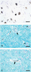
Figure 1
Figure 1
Mercury staining in human brain after exposure to metallic mercury. (A) Mercury in locus ceruleus neurons is seen as small black HgAMG grains, which can readily be distinguished from the pale yellow-brown neuromelanin granules in the cytoplasm. An occasional glial cell (arrow) contains a few HgAMG grains. (B) The cytoplasm of this corticomotor neuron (thick arrow) in the frontal motor cortex contains numerous HgAMG grains. Two nearby small neurons (thin arrows) contain no mercury. The cytoplasm of some glia (asterisks) contains mercury. (C) Mercury staining is seen in the region of this capillary wall (arrow) in the frontal motor cortex. No adjacent small neurons or glia contain mercury. LFB-HgAMG, bar = 50 μm.
Figure 2
Figure 2
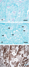
Results of mercury staining in human lower motor neurons and kidney after exposure to metallic mercury. (A) No stainable mercury is present in hypoglossal motor neurons or in nearby glia. LFB-HgAMG, bar = 50 μm. (B) No stainable mercury is present in C3 anterior horn spinal motor neurons (arrows) or surrounding glia. LFB-HgAMG, bar = 50 μm. (C) Heavy mercury staining is present in renal tubules, but not in glomeruli (asterisks). Hematoxylin-HgAMG, bar = 200 μm.
Figure 3
Figure 3
Mercury staining in mouse spinal cord and brain after exposure to mercury. (A) After Hg0 exposure, all large spinal motor neurons in the cervical spinal cord contain HgAMG grains. No glial staining is seen. (B) After Hg0 exposure, all large hypoglossal motor neurons contain HgAMG grains. No glial staining is seen. (C) After Hg0 exposure, dense HgAMG grains are seen in subependymal astrocytes near the ventricle (V), but no locus ceruleus neurons (outlined) contain mercury. (D) After mercuric chloride exposure, no mercury staining is present in layer 5 somatomotor neurons in the frontal lobe, or in surrounding glial cells. Hematoxylin-HgAMG, bar = 50 μm.
Figure 4
Figure 4
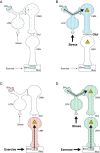
Potential pathways for toxins to enter motor neurons and the possible resulting ALS phenotypes. (A) This circulating toxin is taken up from cerebral (but not peripheral) blood vessels and enters LC neurons. The LC neurons then pass the toxin on to CMNs (thin dashed arrow). With low levels of stress, CMNs take up only a small amount of the toxin, which can readily be handled by cellular detoxifying mechanisms. (B) Under high levels of stress, increased noradrenaline recycling in LC neurons leads to a greater uptake of the toxin into CMNs, which overwhelms the cellular detoxifying mechanisms and could cause an upper motor neuron predominant form of ALS. A toxin that causes CMNs to produce more glutamate will damage LMNs, possibly leading to classical ALS. (C) During strenuous exercise an increased uptake of circulating toxin from intramuscular blood vessels at neuromuscular junctions damages LMNs. This could result in a lower motor neuron predominant form of ALS. (D) This toxin, aided by both stress and exercise, is taken up from both cerebral and intramuscular blood vessels and so can enter CMNs and LMNs. This could result in classical ALS. BVc: cerebral blood vessel, BVm: intramuscular blood vessel, CMN: corticomotor neuron, Glut: glutamate, HgAMG: autometallographic-demonstrable mercury, LC: locus ceruleus, LFB: Luxol-fast blue, Mus: muscle.

Similar articles

Cited by

References

    1. Kiernan MC, Vucic S, Cheah BC, Turner MR, Eisen A, Hardiman O, Burrell JR, Zoing MC. Amyotrophic lateral sclerosis. Lancet. 2011;1:942–955. doi: 10.1016/S0140-6736(10)61156-7. - DOI - PubMed
    1. Chio A, Calvo A, Mazzini L, Cantello R, Mora G, Moglia C, Corrado L, D’Alfonso S, Majounie E, Renton A, Pisano F, Ossola I, Brunetti M, Traynor BJ, Restagno G. Extensive genetics of ALS: a population-based study in italy. Neurology. 2012;1:1983–1989. doi: 10.1212/WNL.0b013e3182735d36. - DOI - PMC - PubMed
    1. Belzil VV, Rouleau GA. Familial ALS: less common than we think? J Neurol Neurosurg Psychiatry. 2012;1:1133–1133. doi: 10.1136/jnnp-2012-303127. - DOI - PubMed
    1. Conte A, Lattante S, Luigetti M, Del Grande A, Romano A, Marcaccio A, Marangi G, Rossini PM, Neri G, Zollino M, Sabatelli M. Classification of familial amyotrophic lateral sclerosis by family history: effects on frequency of genes mutation. J Neurol Neurosurg Psychiatry. 2012;1:1201–1203. doi: 10.1136/jnnp-2012-302897. - DOI - PubMed
    1. Shaw CA, Hoglinger GU. Neurodegenerative diseases: neurotoxins as sufficient etiologic agents? Neuromolecular Med. 2008;1:1–9. doi: 10.1007/s12017-007-8016-8. - DOI - PMC - PubMed

Publication types