

Do brain T2/FLAIR white matter hyperintensities correspond to myelin loss in normal aging? A radiologic-neuropathologic correlation study
- PMID: 24252608
- PMCID: PMC3893472
- DOI: 10.1186/2051-5960-1-14
Do brain T2/FLAIR white matter hyperintensities correspond to myelin loss in normal aging? A radiologic-neuropathologic correlation study
Abstract
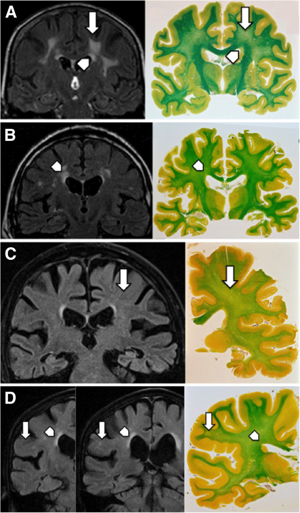
Background: White matter hyperintensities (WMH) lesions on T2/FLAIR brain MRI are frequently seen in healthy elderly people. Whether these radiological lesions correspond to irreversible histological changes is still a matter of debate. We report the radiologic-histopathologic concordance between T2/FLAIR WMHs and neuropathologically confirmed demyelination in the periventricular, perivascular and deep white matter (WM) areas.
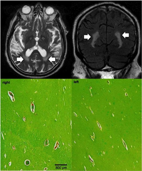
Results: Inter-rater reliability was substantial-almost perfect between neuropathologists (kappa 0.71 - 0.79) and fair-moderate between radiologists (kappa 0.34 - 0.42). Discriminating low versus high lesion scores, radiologic compared to neuropathologic evaluation had sensitivity / specificity of 0.83 / 0.47 for periventricular and 0.44 / 0.88 for deep white matter lesions. T2/FLAIR WMHs overestimate neuropathologically confirmed demyelination in the periventricular (p < 0.001) areas but underestimates it in the deep WM (0 < 0.05). In a subset of 14 cases with prominent perivascular WMH, no corresponding demyelination was found in 12 cases.
Conclusions: MRI T2/FLAIR overestimates periventricular and perivascular lesions compared to histopathologically confirmed demyelination. The relatively high concentration of interstitial water in the periventricular / perivascular regions due to increasing blood-brain-barrier permeability and plasma leakage in brain aging may evoke T2/FLAIR WMH despite relatively mild demyelination.
Figures
References
-
- Ylikoski A, Erkinjuntti T, Raininko R, Sarna S, Sulkava R, Tilvis R. White matter hyperintensities on MRI in the neurologically nondiseased elderly. Analysis of cohorts of consecutive subjects aged 55 to 85 years living at home. Stroke. 1995;1:1171–1177. doi: 10.1161/01.STR.26.7.1171. - DOI - PubMed
MeSH terms
LinkOut - more resources
Full Text Sources
Other Literature Sources
Medical
