

Ribosome biogenesis in skeletal development and the pathogenesis of skeletal disorders
- PMID: 24252615
- PMCID: PMC4020712
- DOI: 10.1016/j.bbadis.2013.11.010
Ribosome biogenesis in skeletal development and the pathogenesis of skeletal disorders
Abstract
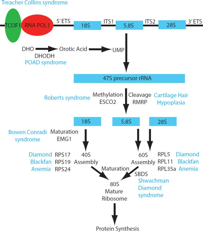
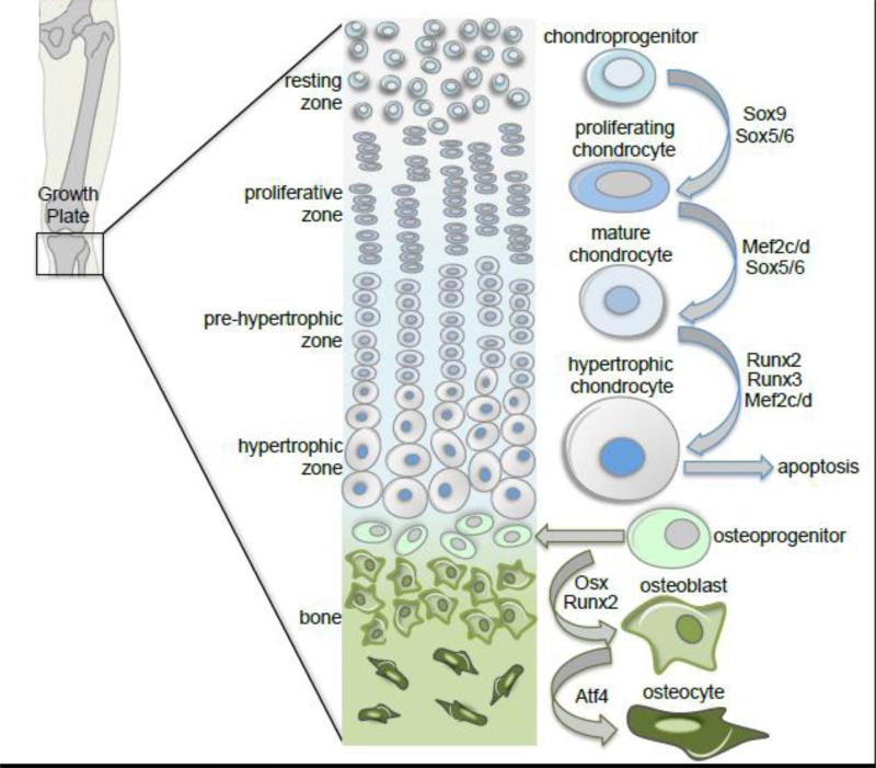
The skeleton affords a framework and structural support for vertebrates, while also facilitating movement, protecting vital organs, and providing a reservoir of minerals and cells for immune system and vascular homeostasis. The mechanical and biological functions of the skeleton are inextricably linked to the size and shape of individual bones, the diversity of which is dependent in part upon differential growth and proliferation. Perturbation of bone development, growth and proliferation, can result in congenital skeletal anomalies, which affect approximately 1 in 3000 live births [1]. Ribosome biogenesis is integral to all cell growth and proliferation through its roles in translating mRNAs and building proteins. Disruption of any steps in the process of ribosome biogenesis can lead to congenital disorders termed ribosomopathies. In this review, we discuss the role of ribosome biogenesis in skeletal development and in the pathogenesis of congenital skeletal anomalies. This article is part of a Special Issue entitled: Role of the Nucleolus in Human Disease.
Keywords: Cartilage hair hypoplasia; Ribosome biogenesis; Roberts syndrome; Shwachman–Diamond syndrome; Skeletal development; Treacher Collins syndrome.
Copyright © 2013 Elsevier B.V. All rights reserved.
Figures
References
-
- Stoll C, Dott B, Roth MP, Alembik Y. Birth prevalence rates of skeletal dysplasias. Clin Genet. 1989;35:88–92. - PubMed
-
- Lafontaine DLJ, Tollervey D. The function and synthesis of ribosomes. Nat Rev Mol Cell Biol. 2001;2:514–520. - PubMed
-
- Kressler D, Hurt E, Baßler J. Driving ribosome assembly. Biochimica et Biophysica Acta (BBA) - Molecular Cell Research. 2010;1803:673–683. - PubMed
-
- Moss T, Stefanovsky V, Langlois F, Gagnon-Kugler T. A new paradigm for the regulation of the mammalian ribosomal RNA genes. Biochem Soc Trans. 2006;34:1079–1081. - PubMed
Publication types
MeSH terms
Substances
Supplementary concepts
Grants and funding
LinkOut - more resources
Full Text Sources
Other Literature Sources
