. 2013 Oct 28:1:71.
doi: 10.1186/2051-5960-1-71.

Lipid accumulation, lipid oxidation, and low plasma levels of acquired antibodies against oxidized lipids associate with degeneration and rupture of the intracranial aneurysm wall

Affiliations

Lipid accumulation, lipid oxidation, and low plasma levels of acquired antibodies against oxidized lipids associate with degeneration and rupture of the intracranial aneurysm wall

Juhana Frösen et al. Acta Neuropathol Commun. .

Abstract

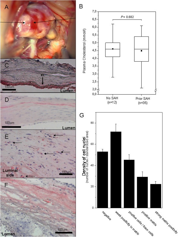
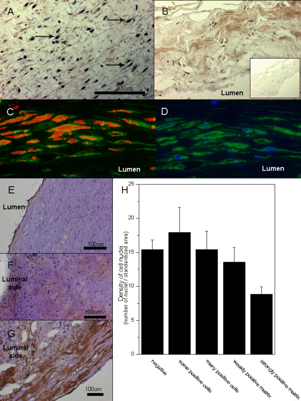
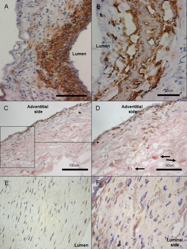
Background: Rupture of a saccular intracranial aneurysm (sIA) causes an often fatal subarachnoid hemorrhage (SAH). Why some sIAs rupture remains unknown. Since sIA walls bear some histological similarities with early atherosclerotic lesions, we hypothesized that accumulation and oxidation of lipids might occur in the sIA wall and might associate with sIA wall degeneration. Tissue samples from sIA fundi (n = 54) were studied with histochemistry and a panel of previously characterized antibodies for epitopes of oxidized LDL (OxLDL). Plasma samples from sIA carriers (n = 125) were studied with ELISA and EIA for IgG and IgM -antibodies against a panel of OxLDL epitopes.

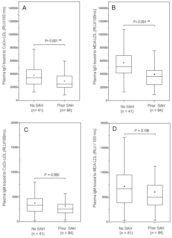
Results: Lipid accumulation, foam cells, and oxidized lipids were found both in unruptured and ruptured sIA walls. Lipid accumulation associated with wall degeneration (P < 0.001), as did the expression of adipophilin, a marker of lipid ingestion by cells. Lipid accumulation associated also with loss of mural cells (P < 0.001), as did the accumulation of OxLDL (P < 0.001). Plasma IgG antibody titers against OxLDL or malondialdehyde modified LDL were higher in patients with unruptured sIAs than in patients with aneurysmal SAH (P ≤ 0.001). A trend but not statistically significant differences were found in plasma IgM antibodies against oxidized lipids.

Conclusions: Accumulation of lipids and their oxidation in the sIA wall associates with the degeneration of the sIA wall. Acquired immunity against oxidized lipid epitopes may be protective of lipid associated sIA wall degeneration, but warrants further studies.


Figures

Figure 1
Figure 1
Atherosclerotic changes, lipid accumulation, and degeneration of the aneurysm wall. Intraoperative view of an MCA aneurysm with atherosclerotic changes (A). Lipids accumulate to the aneurysm wall, despite plasma cholesterol levels that did not significantly differ from “normal” (B, bar graphs display mean and error bars SEM). In non-aneurysmal cerebral artery wall, lipid accumulation is mostly observed subendothelially on the luminal side of the internal elastic lamina (C, sample from a middle cerebral artery, polarized light). In most unruptured sIA walls that show features of mild intimal hyperplasia, lipid accumulation in Oil-Red-O stainings is limited to a matrix layer usually between the outer 1/3 and luminal 2/3 of the wall (D). In aneurysms with more myointimal hyperplasia-like wall, intracellular lipid droplets and foam cells are seen (E). In degenerated and decellularized aneurysm walls, Oil-Red-O staining is mostly extracellular and spread around the degenerated matrix (F). Overall, in the Oil-Red-O stainings the accumulation of lipids was associated with loss of mural cells of the sIA wall (G). The bar graph G displays mean values for density of nuclei (number of nuclei per a standardized surface area) and error bars present SEM.
Figure 2
Figure 2
Accumulation of LDL and oxidized lipids in the aneurysm wall. Intracellular staining pattern for ApoB-100, the core protein of LDL, suggests uptake of cholesterol by cells in the aneurysm wall (A). Staining for ApoB-100 is also observed in the aneurysm wall matrix (B, negative control in the insert). Double stainings (C) for hydroxynonenal (red) and alfa-smooth muscle actin (green) demonstrate accumulation of oxidized lipids in the smooth muscle cells of the aneurysm wall (D, negative control with only alfa-smooth muscle actin in green and nuclei in blue). The accumulation of oxidized LDL (as detected by immunostaining with an antibody against copper oxidized LDL, brown) associates with degeneration of the aneurysm wall (microphotographs: E-G, association with loss of mural cells H). The bar graph H displays mean values for density of nuclei (number of nuclei per a standardized surface area) and error bars present SEM.
Figure 3
Figure 3
Ingestion of lipids by mural cells and inflammatory cells. Adipophilin was expressed in the mural cells of intimal hyperplasia or myointima -like sIA walls (A). In degenerated sIA walls, adipophilin staining does not only stain cells but mostly extracellular debris (B). CD45 and Oil-Red-O double stainings showed presence of inflammatory cells in wall areas abundant in extracellular lipid (C-D, CD45 in brown with DAB, Oil-Red-O and hematoxylin background stain). CD45 positive cells were also found in areas without lipid accumulation (marked with * in C). Most foam cells observed were CD45 negative (marked with arrows in D) and therefore not leukocytes. Expression of 15-lipoxygenase, a known intracellular oxidant of lipids (18), was found in aneurysm wall cells (E, 15-lipoxygenase in brown with DAB and hematoxylin background stain, x40 magnification). Double stainings with CD68 (F, 15-lipoxygenase in brown with DAB and CD68 in blue with AFOS, nuclear fast red background stain) demonstrated that most cells expressing 15-lipoxygenase were not CD68+ macrophages (F), although some double positive cells were also found (data not shown).
Figure 4
Figure 4
Antibodies against oxidized lipids in the plasma. Titers of plasma IgG reactive against oxidized LDL were significantly higher in patients with intracranial aneurysms but no history of SAH (A), as were also the titers of plasma IgG against malondialdehyde (B). Titers for plasma IgM reactive against oxidized LDL (C) or malondialdehyde (D) did not statistically differ among patients with a history of SAH or no history of SAH, although there was a trend towards similar findings as with IgG titers. Box plots represent the median value (horizontal line) and the 25th and 75th percentiles (box edges). Range is given with error bars and the small black box displays mean values.

References

    1. Ingall T, Asplund K, Mahonen M, Bonita R. A multinational comparison of subarachnoid hemorrhage epidemiology in the WHO MONICA stroke study. Stroke. 2000;1:1054–1061. doi: 10.1161/01.STR.31.5.1054. - DOI - PubMed
    1. Broderick JP, Viscoli CM, Brott T, Kernan WN, Brass LM, Feldmann E, Morgenstern LB, Wilterdink JL, Horwitz RI. Major risk factors for aneurysmal subarachnoid hemorrhage in the young are modifiable. Stroke. 2003;1:1375–1381. doi: 10.1161/01.STR.0000074572.91827.F4. - DOI - PubMed
    1. Isaksen J, Egge A, Waterloo K, Romner B, Ingebrigtsen T. Risk factors for aneurysmal subarachnoid haemorrhage: the Tromso study. J Neurol Neurosurg Psychiatry. 2002;1:185–187. doi: 10.1136/jnnp.73.2.185. - DOI - PMC - PubMed
    1. Feigin VL, Rinkel GJ, Lawes CM, Algra A, Bennet DA, Van Gijn J, Anderson CS. Risk factors for subarachnoid hemorrhage: an updated systematic review of epidemiological studies. Stroke. 2005;1:2773–2780. doi: 10.1161/01.STR.0000190838.02954.e8. - DOI - PubMed
    1. Frösen J, Tulamo R, Paetau A, Laaksamo E, Korja M, Laakso A, Niemelä M, Hernesniemi J. Saccular intracranial aneurysm: pathology and mechanisms. Acta Neuropathol. 2012;1:773–786. doi: 10.1007/s00401-011-0939-3. Review. - DOI - PubMed

MeSH terms