Case Reports
. 2013 May 20:1:17.
doi: 10.1186/2051-5960-1-17.

Evidence for new targets and synergistic effect of metronomic celecoxib/fluvastatin combination in pilocytic astrocytoma

Affiliations
Case Reports

Evidence for new targets and synergistic effect of metronomic celecoxib/fluvastatin combination in pilocytic astrocytoma

Sandy Mercurio et al. Acta Neuropathol Commun. .

Abstract

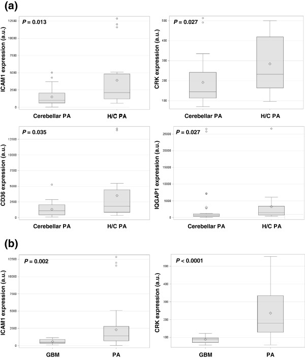
Background: Pilocytic astrocytomas occur predominantly in childhood. In contrast to the posterior fossa location, hypothalamo-chiasmatic pilocytic astrocytomas display a worse prognosis often leading to multiple surgical procedures and/or several lines of chemotherapy and radiotherapy to achieve long-term control. Hypothalamo-chiasmatic pilocytic astrocytomas and cerebellar pilocytic astrocytomas have a distinctive gene signature and several differential expressed genes (ICAM1, CRK, CD36, and IQGAP1) are targets for available drugs: fluvastatin and/or celecoxib.

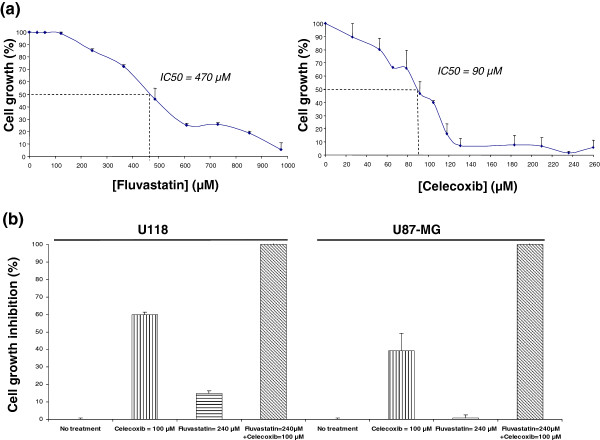
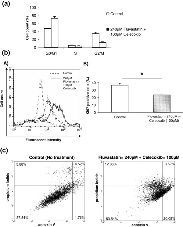
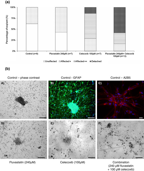
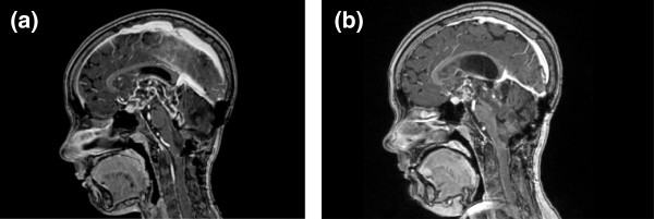
Results: Quantification by RT-Q-PCR of the expression of these genes was performed in a series of 51 pilocytic astrocytomas and 10 glioblastomas: they were all significantly overexpressed in hypothalamo-chiasmatic pilocytic astrocytomas relative to cerebellar pilocytic astrocytomas, and CRK and ICAM1 were significantly overexpressed in pilocytic astrocytomas versus glioblastomas.We used two commercially available glioblastoma cell lines and three pilocytic astrocytoma explant cultures to investigate the effect of celecoxib/fluvastatin alone or in combination. Glioblastoma cell lines were sensitive to both drugs and a combination of 100 μM celecoxib and 240 μM fluvastatin was the most synergistic. This synergistic combination was used on the explant cultures and led to massive cell death of pilocytic astrocytoma cells.As a proof of concept, a patient with a refractory multifocal pilocytic astrocytoma was successfully treated with the fluvastatin/celecoxib combination used for 18 months. It was well tolerated and led to a partial tumor response.

Conclusion: This study reports evidence for new targets and synergistic effect of celecoxib/fluvastatin combination in pilocytic astrocytoma. Because it is non-toxic, this new strategy offers hope for the treatment of patients with refractory pilocytic astrocytoma.


Figures

Figure 1
Figure 1
Expression of transcripts in human tumors. Box plots of expression of transcripts in human tumors show significant differences in (a) ICAM1 (p=0.013), CRK (p=0.027), CD36 (p=0.035) and IQGAP1 (p=0.027) mRNA expression values between hypothalamo-chiasmatic pilocytic astrocytomas (H/C PA) and cerebellar PA and in (b) ICAM1 (p=0.002) and CRK (p<0.0001) mRNA expression values between glioblastomas (GBM) and PA. The lower and upper edges of the box represent the first and third quartile respectively, while a horizontal line within the box indicates the median. The vertical length of the box represents the interquartile range (IQR). The most extreme sample values (within a distance of 1.5 IQR from the median) are the endpoints of the lines extending from the box. a.u.: arbitrary unit.
Figure 2
Figure 2
Cell growth activity measured by MTT assay on U118 cell line treated with drug combination. (a) U118 GBM cell line was cultured with a range of various concentrations of fluvastatin or celecoxib either alone or in combination. After 48 hours, cell growth was measured by MTT assay, and the concentration of each compound that induced 50% growth inhibition (IC50) was determined. U118 cell line was sensitive to both drugs. Results represent the mean of four independent assays plus standard deviation. (b) After 48 hours, cytotoxicity was measured by MTT assay. Fluvastatin (240 μM) potentiates the action of celecoxib (100 μM) on U87-MG and U118 cells causing massive cell growth inhibition in both cell lines (99%) compared to fluvastin used alone (almost none) or celecoxib used alone (50%). Results represent the mean of four independent assays plus standard deviation.
Figure 3
Figure 3
Effect of fluvastatin and celecoxib on U87-MG cell cycle, proliferation and apoptosis. (a) Cells were treated for 24 hours with 240 μM fluvastatin/100 μM celecoxib. Cell cycle of control and treated cells was analyzed by FACS using propidium-iodide-stained nuclei. Percentage of cells in G1, S and G2 phases is shown. (b) Cells were treated for 24 hours with 240 μM fluvastatin/100 μM Celecoxib. Cell proliferation was analyzed by FACS using KI67 staining. (A) Representative experiment of 3 independent experiments is shown. (B) The percentage of KI67-positive cells with mean plus standard deviation of 3 independent experiments is shown. (c) U87-MG cell lines were cultured with fluvastatin (240 μM) or celecoxib (100 μM) either alone or in combination. After 24 hours, cells were collected and analyzed for fluorescein annexin-V and propidium iodide (PI) labelling by FACS in order to distinguish and quantitatively analyze non-apoptotic cells (Annexin-V negative/PI negative, lower-left), early apoptotic cells (Annexin -V positive/PI negative, lower-right), late apoptotic/necrotic cells (Annexin-V positive/PI positive, upper-right) and dead cells (Annexin V negative/PI positive, upper-left). This double-labelling was performed on untreated cells and treated cells with the drug combination (100 μM of celecoxib/240 μM of fluvastatin).
Figure 4
Figure 4
In vitro analysis of the cytotoxicity of fluvastatin and/or celecoxib on PA explant cultures. PA explants were grown in DMEM 10% FCS for 10 days and then treated with fluvastatin (240 μM) or celecoxib (100 μM) either alone or in combination. Untreated explants were used as controls. (a) Regarding the 4 level scale that we have established (“unaffected”, “affected +”, “affected ++”, “detached”), we recorded the percentage of explants in each state for each condition in PA-PET explant culture. (b) Ten days after explantation, cell growth was observed around the explants and in untreated conditions, explants and cell growth around the explants were “unaffected” (A). Expression of GFAP (B) and A2B5 (C) was analyzed by immunofluorescent staining to confirm the glial nature of cultured cells. When used alone, 240 μM fluvastatin treatment had little effect on explants but most cells around them mainly became round and lost their adhesion (D, “affected +” state). Celecoxib treatment (100 μM) mainly induced the “affected ++” state: damage to both explants and cell growth (E). Explants treated with the combination were totally disrupted and scattered (F, “detached” state). Scale bar: 100 μm.
Figure 5
Figure 5
Cerebral MRI, sagittal sections, T1 weighted with gadolinium injection. (a) May 2009: Before antitumoral treatment with celecoxib/fluvastatin. Hypersignal in the hypothalamo-chiasmatic region, fourth ventricle, cerebellum. Post-operative reshuffle of the posterior fossa. (b) September 2010: After 16 months of antitumoral treatment with celecoxib/fluvastatin. Decrease in contrast enhancement of the hypothalamo-chiasmatic, fourth ventricle and cerebellum lesions.

Similar articles

Cited by

References

    1. Fernandez C, Figarella-Branger D, Girard N, Bouvier-Labit C, Gouvernet J. Paz Paredes A, Lena G: Pilocytic astrocytomas in children: prognostic factors–a retrospective study of 80 cases. Neurosurgery. 2003;1:544–553. doi: 10.1227/01.NEU.0000079330.01541.6E. discussion 554–545. - DOI - PubMed
    1. Qaddoumi I, Sultan I, Gajjar A. Outcome and prognostic features in pediatric gliomas: a review of 6212 cases from the Surveillance, Epidemiology, and End Results database. Cancer. 2009;1:5761–5770. doi: 10.1002/cncr.24663. - DOI - PMC - PubMed
    1. Tihan T, Ersen A, Qaddoumi I, Sughayer MA, Tolunay S, Al-Hussaini M, Phillips J, Gupta N, Goldhoff P, Baneerjee A. Pathologic characteristics of pediatric intracranial pilocytic astrocytomas and their impact on outcome in 3 countries: a multi-institutional study. Am J Surg Pathol. 2011;1:43–55. - PubMed
    1. Cin H, Meyer C, Herr R, Janzarik WG, Lambert S, Jones DT, Jacob K, Benner A, Witt H, Remke M. Oncogenic FAM131B-BRAF fusion resulting from 7q34 deletion comprises an alternative mechanism of MAPK pathway activation in pilocytic astrocytoma. Acta Neuropathol. 2011;1:763–774. doi: 10.1007/s00401-011-0817-z. - DOI - PubMed
    1. Hawkins C, Walker E, Mohamed N, Zhang C, Jacob K, Shirinian M, Alon N, Kahn D, Fried I, Scheinemann K. BRAF-KIAA1549 fusion predicts better clinical outcome in pediatric low-grade astrocytoma. Clin Cancer Res. 2011;1:4790–4798. doi: 10.1158/1078-0432.CCR-11-0034. - DOI - PubMed

Publication types

MeSH terms