Case Reports
. 2013 Nov 11:1:72.
doi: 10.1186/2051-5960-1-72.

Rapidly progressive dementia with thalamic degeneration and peculiar cortical prion protein immunoreactivity, but absence of proteinase K resistant PrP: a new disease entity?

Affiliations
Case Reports

Rapidly progressive dementia with thalamic degeneration and peculiar cortical prion protein immunoreactivity, but absence of proteinase K resistant PrP: a new disease entity?

Gabor G Kovacs et al. Acta Neuropathol Commun. .

Abstract

Background: Human prion diseases are a group of rare fatal neurodegenerative conditions with well-developed clinical and neuropathological diagnostic criteria. Recent observations have expanded the spectrum of prion diseases beyond the classically recognized forms.

Results: In the present study we report six patients with a novel, apparently sporadic disease characterised by thalamic degeneration and rapidly progressive dementia (duration of illness 2-12 months; age at death: 55-81 years). Light and electron microscopic immunostaining for the prion protein (PrP) revealed a peculiar intraneuritic distribution in neocortical regions. Proteinase K resistant PrP (PrPres) was undetectable by Western blotting in frontal cortex from the three cases with frozen tissue, even after enrichment for PrPres by centrifugation or by phosphotungstic acid precipitation. Conformation-dependent immunoassay analysis using a range of PK digestion conditions (and no PK digestion) produced only very limited evidence of meaningful D-N (denatured/native) values, indicative of the presence of disease-associated PrP (PrPSc) in these cases, when the results were compared with appropriate negative control groups.

Conclusions: Our observation expands the spectrum of conditions associated with rapidly progressive dementia and may have implications for the understanding of the pathogenesis of prion diseases.


Figures

Figure 1
Figure 1
Neuropathological observations. Prominent neuronal loss and gliosis in the medial thalamus. (a: H&E, b: GFAP; representative image of case 1) associated with segmental gliosis and vacuolation in the inferior olives (c; representative image of case 5; inset demonstrates vacuolated neuron). Moderate gliosis mainly in the upper layers of the cortex (d; representative image of case 4). Bar in a indicates 50 μm for a, 30 μm for b, 100 μm for c, and 150 μm for d.
Figure 2
Figure 2
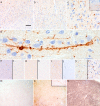
Immunostaining for PrP. Immunostaining for PrP revealed tiny granular deposits depicting neuronal processes in a linear or worm-like appearance predominantly in the 2nd and 3rd layers in neocortical regions. (a-c; representative images of case 2; right upper inset in c shows similar immunoreactivity in the cerebellum in case 3). Occasionally, small cell bodies were depicted as well (d). These were detectable also by antibodies 6H4 (e), and KG9 (f), only focally by 3F4 (g), and not by the anti-N-terminal BG4 antibody (h all representative images of case 4). The PrP immunoreactivity (i) was abolished after PK treatment (j; temporal cortex of case 1), while it was slightly more detectable when using only elongated citrate buffer pretreatment and omitting formic acid pretreatment (k: with, and l: without formic acid, represented by the occipital cortex of case 5). PET-blot examination performed using Thermolysin digested PrPC completely from the frontal cortex brain tissue of a non-CJD control case (m), while in the present cases the worm-like structures (enlarged in right upper inset) remained visible (n), and in the cortex of a subject with CJD abundant PrPSc deposition was seen (o). Bar in a represents 150 μm for a, 50 μm for b, e-l, 30 μm for c, 10 μm for d, and 400 μm for m-o.
Figure 3
Figure 3
Ultrastructural localisation of PrP using antibody 12F10 in the same frontal cortex section where light microscopical immunohistochemistry revealed linear and worm-like immunoreactivity. Panel a-d demonstrates immunogold labeling specific for PrP mainly associated with endosome-like structures in the pericaryon (indicated by arrows; Nu: nucleus). Intraneuritic accumulation of PrP in longitudinal (e) and cross (f) sections of myelinated axons. Measuring bars represent 2 μm in a, c, d, and e, and 500 nm in b and f.
Figure 4
Figure 4
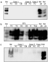
Western blot analysis of present cases 1, 2, and 4 and an Austrian FFI case using 3F4 as the primary antibody. PrPres type 1, 2A and 2B standards were run in the lanes as indicated. a: PrPres was undetectable in 200 μl (of 10% wt/vol brain homogenate) after centrifugal concentration. b: Centrifugal concentration (100 μl of 10% wt/vol brain homogenate) shows readily detectable PrPres in the A-FFI case. c: PrPres was undetectable in 500 μl of 10% wt/vol brain homogenate from cases 1, 2 and 4 after NaPTA precipitation. FC, frontal cortex; OC, occipital cortex; CB, cerebellum; TC, temporal cortex; H, hippocampus; Thal, thalamus.
Figure 5
Figure 5
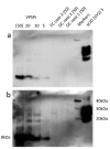
Western blot analysis of PrPres in frontal cortex (FC) or occipital cortex (OC) of cases 1 and 2 compared to cortical samples of a variably protease sensitive prionopathy (VPSPr) case and a sporadic Creutzfeldt-Jakob disease of the VV2 subtype (sCJD (VV2)). Sample loadings (μl of a 10% w/v brain homogenate) are shown for all lanes. Sample loading in square brackets ([]) denote volumes concentrated prior to loading. The positions molecular mass of marker proteins (Markers) and the low molecular mass PrPres characteristic of VPSPr are given in kilodaltons (kDa). (a) and (b) show short (3 minutes) and long (30 minutes) exposures of the same Western blot.
Figure 6
Figure 6
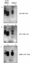
Western blot analysis of grey matter enriched frontal cortex from a sCJD MM1 patient and case 2 using anti-PrP antibodies 1E4. (a), 3F4 (b) and 94B4 (c) (epitopes as indicated on the figure). Non-PK treated samples (−) were loaded at 6 μl/lane of 10% w/v brain homogenate, whereas PK-treated samples (+) were loaded at the maximum of 24 μl/lane of 10% w/v brain homogenate, but without any sample concentration pre-treatment.
Figure 7
Figure 7
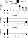
CDI analysis of the frontal cerebral cortices of the case 1, 2 and 4, and an FFI case after treatment with 0, 2.5 and 50 μg/ml PK. (panels a, b and c, respectively). Frontal cortical brain homogenates from cases of sporadic Creutzfeldt-Jakob disease (sCJD) types MM1 and MM2T (the latter also known as sporadic fatal insomnia, sFI) were also analysed for comparison. Note that the range of the y-axis decreases from panel a to b to c. For case 1 and 2 duplicate frozen samples, labeled ‘a’ and ‘b’, were available for CDI analysis.
Figure 8
Figure 8
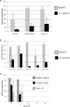
CDI analysis of cases compared with negative control groups. (a) Comparison of CDI values (means) for non-CJD control, sudden death control and cerebral cortex samples. Frontal cortex samples from ten cases from the Medical Research Council Edinburgh Brain and Tissue bank were analysed by CDI as controls. Five of these were Sudden Death cases with no history of a neurological condition (‘Sudden death’), whereas another five were cases initially referred to NCJDRSU as suspected CJD cases, but given an alternative final diagnosis (‘non-CJD’). These samples were analysed in comparison with three samples of cerebral cortex from cases 1 (one sample) and 2 (two samples). The upper and lower dashed lines indicate the D-N cut-off values, these being the [mean + (2.5 X Standard Deviation)] for sudden death and non-CJD cerebral cortex tissue, respectively. (b) Comparison of CDI values (means) for non-CJD control and sudden death control versus the individual samples from cases 1 and 2. (c) The D-N values are shown from CDI analysis of a sample of frontal cortex from cases 1 and 2 (striped bar) and sudden death cases (black bar) or non-CJD neurological control cases (grey bars), following treatment with various concentrations of PK as indicated on the x-axis.

Similar articles

Cited by

References

    1. Head MW, Ironside JW. Review: Creutzfeldt-Jakob disease: prion protein type, disease phenotype and agent strain. Neuropathol Appl Neurobiol. 2012;38(4):296–310. doi: 10.1111/j.1365-2990.2012.01265.x. - DOI - PubMed
    1. Prusiner SB. Prions. Proc Natl Acad Sci USA. 1998;95(23):13363–13383. doi: 10.1073/pnas.95.23.13363. - DOI - PMC - PubMed
    1. Parchi P, Castellani R, Capellari S, Ghetti B, Young K, Chen SG, Farlow M, Dickson DW, Sima AA, Trojanowski JQ. et al.Molecular basis of phenotypic variability in sporadic Creutzfeldt-Jakob disease. Ann Neurol. 1996;39(6):767–778. doi: 10.1002/ana.410390613. - DOI - PubMed
    1. Piccardo P, Dlouhy SR, Lievens PM, Young K, Bird TD, Nochlin D, Dickson DW, Vinters HV, Zimmerman TR, Mackenzie IR. et al.Phenotypic variability of Gerstmann-Straussler-Scheinker disease is associated with prion protein heterogeneity. J Neuropathol Exp Neurol. 1998;57(10):979–988. doi: 10.1097/00005072-199810000-00010. - DOI - PubMed
    1. Parchi P, Strammiello R, Giese A, Kretzschmar H. Phenotypic variability of sporadic human prion disease and its molecular basis: past, present, and future. Acta Neuropathol. 2011;121(1):91–112. doi: 10.1007/s00401-010-0779-6. - DOI - PubMed

Publication types