. 2013 Nov 12:1:73.
doi: 10.1186/2051-5960-1-73.

In vivo modification of Abeta plaque toxicity as a novel neuroprotective lithium-mediated therapy for Alzheimer's disease pathology

Affiliations

In vivo modification of Abeta plaque toxicity as a novel neuroprotective lithium-mediated therapy for Alzheimer's disease pathology

Laura Trujillo-Estrada et al. Acta Neuropathol Commun. .

Abstract

Background: Alzheimer's disease (AD) is characterized by the abnormal accumulation of extracellular beta-amyloid (Abeta) plaques, intracellular hyperphosphorylated tau, progressive synaptic alterations, axonal dystrophies, neuronal loss and the deterioration of cognitive capabilities of patients. However, no effective disease-modifying treatment has been yet developed. In this work we have evaluated whether chronic lithium treatment could ameliorate the neuropathology evolution of our well characterized PS1M146LxAPPSwe-London mice model.

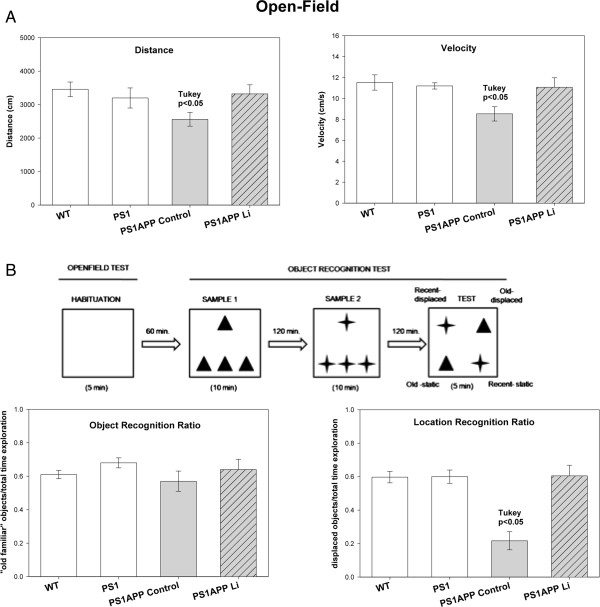
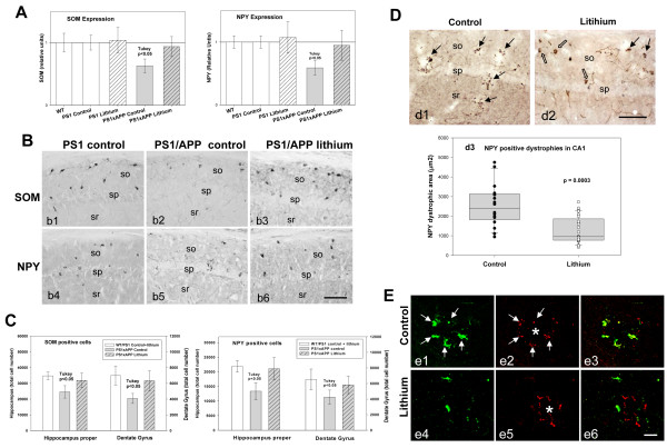
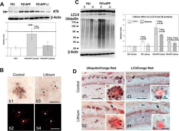
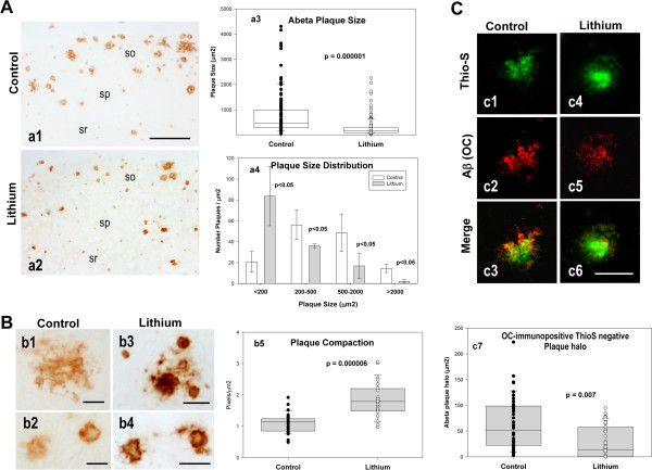
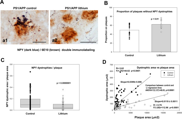
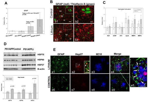
Results: Though beneficial effects of lithium have been previously described in different AD models, here we report a novel in vivo action of this compound that efficiently ameliorated AD-like pathology progression and rescued memory impairments by reducing the toxicity of Abeta plaques. Transgenic PS1M146LxAPPSwe-London mice, treated before the pathology onset, developed smaller plaques characterized by higher Abeta compaction, reduced oligomeric-positive halo and therefore with attenuated capacity to induce neuronal damage. Importantly, neuronal loss in hippocampus and entorhinal cortex was fully prevented. Our data also demonstrated that the axonal dystrophic area associated with lithium-modified plaques was highly reduced. Moreover, a significant lower accumulation of phospho-tau, LC3-II and ubiquitinated proteins was detected in treated mice. Our study highlights that this switch of plaque quality by lithium could be mediated by astrocyte activation and the release of heat shock proteins, which concentrate in the core of the plaques.

Conclusions: Our data demonstrate that the pharmacological in vivo modulation of the extracellular Abeta plaque compaction/toxicity is indeed possible and, in addition, might constitute a novel promising and innovative approach to develop a disease-modifying therapeutic intervention against AD.


Figures

Figure 1
Figure 1
Lithium treatment improved the behavioral/memory deficiencies of PS1xAPP mice. (A) Open-field test. One-way ANOVA revealed significant differences between groups in both distance (F(3,24) = 6.33; p = 0.006) and velocity F(3,24) = 8.02, p = 0.002). Post-hoc analysis showed significant differences in PS1xAPP control compared with WT/PS1 and PS1xAPP lithium (p < 0.05). (B) Behavioral protocol and memory measures. Sixty min after habituation to the open-field, animals received three samples trials with 120 min inter-trial as depicted. Each symbol (triangle and star) represents one type of object. One-way ANOVA F(3,24) = 21.15 p = 0.0001) revealed significant differences between groups in place memory index. Post-hoc analysis showed significant difference in PS1xAPP control compared with WT/PS1 controls and PS1xAPP lithium.
Figure 2
Figure 2
Lithium treatment avoided neuronal loss in the hippocampus of PS1xAPP mice. (A) The expression of SOM and NPY (n= 20 mice per group) was significantly lower in PS1xAPP control group (ANOVA F(4,96) = 49.63, p = 0.00001. Tukey p < 0.05 or F(4,96) = 27.24, p = 0.0001, Tukey p < 0.05 for SOM and NPY respectively). WT, PS1 (control or lithium) and PS1xAPP lithium displayed no differences. B) SOM (b1-b3) or NPY (b4-b6) immunolabeled sections through CA1 subfield of PS1 control or PS1xAPP control and lithium groups. C) Total SOM or NPY cell number in hippocampus proper and dentate gyrus was assessed by stereology (n = 5 per genotype and treatment). WT and PS1 groups displayed no differences and data were pooled. Only control PS1xAPP displayed a significant reduction in SOM (ANOVA F(2,22) = 14.57, p = 0.00002 or F(2.22) = 11.84, p = 0.0008 for hippocampus proper and dentate gyrus, respectively) and NPY (ANOVA F(2,22) = 17.02, p = 0.0001 or F(2,22) = 6.87, p = 0.008 for hippocampus proper and dentate gyrus, respectively) cell number. D) NPY-positive dystrophies (black arrows) in CA1 subfield of PS1xAPP control (d1) and lithium mice (d2) (open arrows: NPY cell bodies). (d3) Quantitative analysis (n = 4 mice per group; 6 sections per animal) demonstrated the existence of a significant reduction of NPY-positive dystrophic area (μm2) in PS1xAPP lithium. E) Co-localization of SOM (green) and AT8 (red) in dystrophies assessed using confocal microscopy. In PS1xAPP control mice (e1 to e3), SOM positive dystrophies (arrows), surrounding Abeta plaque (asterisk), were positive for AT8. Lithium produced a reduction in both SOM (e4) and AT8 (e5) positive dystrophies and in the co-localization between SOM and AT8 in dystrophies (e6). Scale bars: b1-b6 100 μm; d1 and d2 100 μm; e1-e6 20 μm.
Figure 3
Figure 3
Lithium treatment reduced the dystrophic pathology associated to Abeta plaques. A) Representative western blot (n = 7 mice per group) and quantitative analysis of phosphorylated tau, determined using AT8 clone, of total proteins from PS1 control, PS1xAPP control and PS1xAPP lithium-treated mice. For quantification, the AT8 levels were referred to PS1 control group. The AT8 levels were significantly increased in PS1xAPP control mice (ANOVA F(2,18) = 22.66, p = 0.0001; Tukey p < 0.05) whereas PS1 control and PS1xAPP lithium displayed no differences. B) Representative AT8 positive dystrophies surrounding Abeta plaques from control (b1 and b2) or lithium treated (b3 and b4) PS1xAPP mice. C) Representative western blots and quantitative analysis of steady-state levels of LC3-II and ubiquitinated proteins in PS1 and PS1xAPP control and treated mice (n = 4 per genotype and treatment). A clear and significant accumulation of both LC3-II (ANOVA F(3,12) = 32.52, p = 0.001) and ubiquitinated proteins (ANOVA (F(3,12) = 63.67, p = 0.00001) was observed in PS1xAPP control mice. Lithium treatment reversed this pathology. The post-hoc analysis, using Tukey test, was indicated in the figure. D) Ubiquitin (d1 and d2) and LC3 (d3 and d4) immunolabeled hippocampal sections (counterstained with Congo red for Abeta plaques) of control and lithium treated PS1xAPP mice corroborated the accumulation of both markers, associated with dystrophic neurites surrounding amyloid plaques (d1 and d3) and the patent lithium reduction (d2 and d4) in the presence of ubiquitin and LC3 positive dystrophies. Inserts in d1-d4 showed higher magnification details of the dystrophies surrounding Abeta plaques. so, stratum oriens; sp, stratum pyramidale; sr, stratum radiatum. Scale bars: b1, b3 50 μm; b2, b4 100 μm; d1 to d4 200μm; inserts 25 μm.
Figure 4
Figure 4
Lithium treatment reduced the size and increased the compaction of the extracellular Abeta plaques. A) Lithium treatment reduced the size of Abeta deposits. Plaques were immunostained with anti-Abeta42 antibody. Representative images (a1 and a2) and quantitative analysis (a3 and a4) of Abeta plaque size from the CA1 subfield of control and lithium-treated PS1xAPP mice. (a3) The individual plaque size (μm2) was quantitatively assessed from 25 sections of 6 different control or lithium treated PS1xAPP mice. (a4) The plaque size distribution was determined by calculating the number of plaques falling into distinct area categories (ranging from <200 μm2 to >2000 μm2). For each category, the difference between control and lithium was determined by t-test. so, stratum oriens; sp, stratum pyramidale; sr, stratum radiatum. B) Lithium treatment increased the Abeta plaque compaction. Signal density of Abeta42 immunostained plaques from control (b1, b2) and lithium-treated (b3, b4) mice were measured in the CA1 subfield of hippocampus. Optical densities (pixel/μm2) of plaques from 5 sections/mouse and 5 mice per group was represented in the graph. C) Plaque oligomeric halo, considered as the difference between OC- and Thio-S-stained areas was reduced by lithium. Plaques were sequentially staining with Thio-S and the OC antibody. (c1-c6) Representative images of a similar size plaque in the CA1 region of control (c1-c3) or lithium-treated (c4-c6) mice showing the plaque halo. (c7) Similar size plaques (< 200 μm2) were analyzed (15 plaques per animal, n = 3 mice per group). Scale bars: a1, a2 200 μm; b1-b4 25 μm; c1-c6 20 μm.
Figure 5
Figure 5
Lithium treatment decreased the Abeta toxicity. A) NPY-labeled dystrophic neurites, surrounding 6E10-labeled Abeta plaques, were compared in CA1 field of hippocampus from control (a1) and lithium-treated (a2) PS1xAPP mice (5 sections per mice, 3 mice per group). B) Lithium treatment increased the proportion of Abeta plaques displaying no apparent NPY-dystrophies. The difference between groups (indicated in the figure) was assessed using t-test. C) Lithium also significantly (using non-parametric Wilcoxon test) reduced the NPY dystrophic area, normalized by the corresponding plaque area. D) The decrease on Abeta toxicity, at all plaque sizes, was also assessed by plotting the individual NPY dystrophic area versus Abeta plaque area. The individual data from both groups were fitted to a linear model and the possible difference between groups was analyzed using Multiple Regression Analysis. As shown, both groups (control and lithium) could be fitted to a linear model and both regression lines were statistically different (ANOVA F(3,145) = 38.00; p = 0.00001). Furthermore, the slopes of the fitted data were also significantly different between both groups (Conditional Sum of Squares, see Results). Scale bars: a1 25 μm, a2 50 μm.
Figure 6
Figure 6
Lithium increased the astrocyte activation and the Hsps levels. A-B) Lithium treatment increased the astrocyte activation assessed by qPCR (A) and GFAP immunoreactivity (B). GFAP expression (A) was assayed in hippocampal samples from 10 mice per group. GFAP expression increased in PS1xAPP control group and further increased in PS1xAPP lithium-treated mice. ANOVA (F(3,36) = 18.82, p = 0.0001). The post-hoc analysis using Tukey is presented in the graph. B) Representative confocal images through the CA1 field of control (b1, b2) and lithium-treated (b3, b4) mice, showing GFAP-immunoreactive astrocytes (red) and Thioflavin-stained Abeta plaques (green). As shown, GFAP immunoreactivity is clearly higher in lithium-treated mice. C) Lithium did not modify the expression of NGF,GDNF, NT-5, MMP9, MMP3 or ApoE, determined by qPCR (n = 4 mice per group). D) Lithium treatment increased the levels of three Hsps (Hsp70, Hsp60 and Hsp27). Representative western blots and quantitative analysis of Hsps using protein extract from hippocampus of control and lithium-treated PS1xAPP mice (n = 4 mice per group). The expression of all three Hsps was normalized using PS1 control (not shown), and the significance was determined by t-test. E) Confocal images through the CA1 subfield showing GFAP/Hsp27/Abeta triple immunofluorescence labeling. In lithium-treated mice (e1-e5), the core of the Abeta plaques (labeled by 6E10, in blue) is intensely stained with the anti-Hsp27 antibody (in red), as well as astrocytic puncta processes (labeled by GFAP, in green) surrounding the plaques (see insert in e2 showing double GFAP-Hsp27 labeling, and the triple labeling detail in e5). On the contrary, in control mice (e6-e9), the core of the Abeta plaques appears almost devoid of Hsp27 immunostaining and low or no immunoreactivity was observed in astrocytes surrounding the plaques. Scale bars, d1-d9 20 μm, insert in a2 10 μm.

References

    1. Hardy J, Selkoe DJ. The Amyloid hypothesis of Alzheimer’s disease: progress and problems on the road to therapeutics. Science. 2002;297:353–356. doi: 10.1126/science.1072994. - DOI - PubMed
    1. Wyss-Coray T, Rogers J. Inflammation in Alzheimer Disease’s. A Brief Review of the Basic Science and Clinical Literature. Cold Spring Harb Perspect Med. 2012;2:a006346. doi: 10.1101/cshperspect.a006346. - DOI - PMC - PubMed
    1. Diniz B, Machado-Vieira R, Forlenza O. Lithium and neuroprotection: translational evidence and implications for the treatment of neuropsychiatric disorders. Neuropsychiatr Dis Treat. 2013;9:493–500. - PMC - PubMed
    1. Forlenza O, Paula V, Machado-Vieira R, Diniz B, Gattaz W. Does Lithium Prevent Alzheimer’s disease? Drugs Aging. 2012;29:335–342. doi: 10.2165/11599180-000000000-00000. - DOI - PubMed
    1. Nunes PV, Forlenza OV, Gattaz WF. Lithium and risk for Alzheimer’s disease in elderly patients with bipolar disorder. B J Psychiatry. 2007;190:359–360. doi: 10.1192/bjp.bp.106.029868. - DOI - PubMed

Publication types

MeSH terms