. 2014 Jan 15;385(2):200-10.
doi: 10.1016/j.ydbio.2013.11.009. Epub 2013 Nov 16.

Cochleovestibular nerve development is integrated with migratory neural crest cells

Affiliations

Cochleovestibular nerve development is integrated with migratory neural crest cells

Lisa L Sandell et al. Dev Biol. .

Abstract

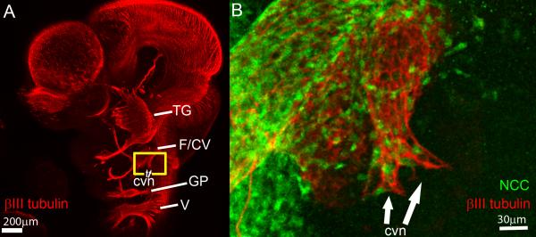
The cochleovestibular (CV) nerve, which connects the inner ear to the brain, is the nerve that enables the senses of hearing and balance. The aim of this study was to document the morphological development of the mouse CV nerve with respect to the two embryonic cells types that produce it, specifically, the otic vesicle-derived progenitors that give rise to neurons, and the neural crest cell (NCC) progenitors that give rise to glia. Otic tissues of mouse embryos carrying NCC lineage reporter transgenes were whole mount immunostained to identify neurons and NCC. Serial optical sections were collected by confocal microscopy and were compiled to render the three dimensional (3D) structure of the developing CV nerve. Spatial organization of the NCC and developing neurons suggest that neuronal and glial populations of the CV nerve develop in tandem from early stages of nerve formation. NCC form a sheath surrounding the CV ganglia and central axons. NCC are also closely associated with neurites projecting peripherally during formation of the vestibular and cochlear nerves. Physical ablation of NCC in chick embryos demonstrates that survival or regeneration of even a few individual NCC from ectopic positions in the hindbrain results in central projection of axons precisely following ectopic pathways made by regenerating NCC.

Keywords: 3-dimensional; 3D; Axon; CV; Cochlea; Craniofacial; E; Ear; GFP; Green Fluorescent Protein; NCC; Neural crest; Neuron; Otic; P; R; Vestibular; cochleovestibular; embryonic day; neural crest cells; postnatal day; rhombomere.


Figures

Figure 1
Figure 1. Mouse Cre driver;reporter combinations used to visualize NCC in CV nerve development
(A and C) Embryos carrying Wnt1Cre;R26R (A) or Wnt1Cre;Z/EG (C) have all NCC and derivatives marked by expression of lacZ or GFP, respectively. Single arrowhead indicates second pharyngeal arch with all NCC labeled by Wnt1Cre-mediated recombination. (B and D) Within the cranial region, embryos carrying 6.5Pax3Cre;R26R (B) or 6.5Pax3Cre;Z/EG (D) preferentially label NCC emanating from R4 with β-galactosidase activity or GFP, respectively. The R4 NCC labeling is mosaic. Double arrowhead indicates second pharyngeal arch with mosaic labeling of R4 NCC. Hypaxial muscle progenitors are also labeled by this Cre driver (not shown). (A and B) Staining for β-galactosidase activity reveals NCC and derivatives labeled by Cre mediated recombination. Color image of β-galactosidase signal is overlain onto grayscale image of fluorescent DAPI signal to emphasize embryo morphology. (C-D) Double immunostain for GFP (green)and neuronal βIII tubulin (red) reveals NCC and neurons. DAPI labeling of all cell nuclei reveals pharyngeal arch morphology. Yellow box in (A) indicates approximate region of images shown in panels (C) and (D). Scale bar in (C) and (D) = 150μm.
Figure 2
Figure 2. NCC stream co-localizes with nested facial and CV ganglia in E10.5 mouse embryos
(A) Whole embryo stained with DAPI and imaged on fluorescent stereo microscope. Yellow box corresponds to region of imaged in (2B and 3A). (B) Wnt1Cre;Z/EG embryo double immunostained with GFP for NCC and ISL1 for neuronal cell bodies. DAPI stain indicates all cell nuclei. Nested facial and CV ganglia are visible anterior to the otic vesicle. Stream of NCC and derivatives emanates from R4, surrounds the nested ganglia, and fills the second pharyngeal arch. (C) NCC stream emanating from R4 envelopes nested facial and CV ganglion neurons as a sleeve. (D) Rare NCC are visible within the aggregated neuronal cell bodies of the CV ganglion, primarily near regions of the otic vesicle with low level ISL1 signal indicative of delaminating neuroblasts. Dotted lines in (C-D) indicate epithelium of otic vesicle. Arrowhead, region of otic vesicle of delaminating neuroblasts indicated by low level ISL1 signal; dotted lines, otic vesicle epithelium; CVG, cochleovestibular ganglion; FG, facial ganglion; HB, hindbrain; ovl, otic vesicle lumen; PA2, pharyngeal arch 2.
Figure 3
Figure 3. Early CV ganglion resembles a saddle straddling ventral otic vesicle
Solid surface rendering of ganglion fluorescence signal and optical cropping of region of interest allows visualization of CV ganglion morphology of E10.5 embryo. ISL1 fluorescence (neuronal cell bodies) is rendered as solid surface to allow visualization of E10.5 mouse CV ganglion. (A) Otic region as indicated by yellow box in (Fig. 2A). Solid surface rendering of ISL1 signal indicates nested facial and CV ganglion straddle ventral otic vesicle at proximal region of second pharyngeal arch. Uncropped GFP immunostain of Wnt1Cre;Z/EG signal reveals NCC and derivatives filling the first and second pharyngeal arches. DAPI signal is cropped to define position of otic vesicle epithelium. (B) Lateral view of surface-rendered ganglion surfaces reveal two prongs extending across ventral otic vesicle. (C-C’) Ventral view of rendered ganglia cell bodies reveals nested organization of facial and CV ganglia and ventral prongs of CV ganglion that extend medially and laterally. (D-D’) Dorsal view of surface rendered ganglia reveals prongs of CV ganglion. Scale bars = 100μm with respect to individual image planes, CVG, CV ganglion; FG, facial ganglion; HB, hindbrain; lp, lateral projection; mp, medial projection; OV, otic vesicle; PA1, pharyngeal arch 1; PA2, pharyngeal arch 2.
Figure 4
Figure 4. CV Axons and neurites form a lattice with NCC intercalated in interstitial spaces at early stages of CV nerve development
(A) Whole mount E10.5 mouse head stained with βIII neuronal tubulin to visualize central axons and peripheral neurites of cranial nerves. Yellow box indicates region imaged in panel B. (B) CV nerve of Wnt1Cre;Z/EG embryo immunostained for βIII neuronal tubulin (neurons) and GFP (NCC) indicates peripheral vestibular neurites are organized as a lattice with NCC intercalated between the spaces of the lattice and at distal-most extent of neurite projections . cvn, terminal peripheral neurites of CV nerve; F/CV, facial nerve/CV nerve; GP, glossopharyngeal nerve, TG, trigeminal nerve; V, vagal nerve.
Figure 5
Figure 5. Progression of CV nerve neurite outgrowth integrated with migratory NCC
(A-D) Lateral view of CV nerve and otic vesicle morphology visualized in isolation by optical cropping of individual image planes in confocal image stacks. Whole embryos or isolated inner ears were immunostained for GFP to identify NCC, and with βIII tubulin to identify central axons and peripheral neurites. DAPI staining reveals all cell nuclei within optically cropped otic vesicle. (A) whole CV nerve and otic vesicle of E10.5 Wnt1CRE;Z/EG, (B) whole CV nerve and otic vesicle of E11.5 6.5Pax3Cre;ZEG, (C) whole CV nerve and developing inner ear of E12.5 6.5Pax3Cre;Z/EG, (D) developing cochlea and fan of neurites from early spiral nerve of E12.5 6.5Pax3Cre;Z/EG with accompanying population of NCC at region of extending peripheral neurites. (E) Schematic representation of E12.5 CV nerve and otic vesicle as in panel (C) to aid in visualization of 3D organization. (F) β-galactosidase staining of inner ear of neonatal 6.5Pax3Cre;R26R pup reveals NCC evenly distributed throughout the CV nerve. Scale bars = 100μm with respect to individual image planes. aa, anterior ampulla; aco, apical cochlea; ax, axonal projection to hindbrain; bco, basal cochlea; cn, cochlear nerve; cvg, cochleovestibular ganglion; dov, dorsal otic vesicle; hb, direction of hindbrain; ivn, inferior vestibular nerve; la, lateral ampulla; pa, posterior ampulla; pro, projecting cochlear neurites; sac, saccule; sg, spiral ganglion; svn, superior vestibular nerve; ut, utricle; vg, vestibular ganglia; vov, ventral otic vesicle.
Figure 6
Figure 6. Elimination of NCC disrupts outgrowth of central axons
Test of chick CV nerve development following ablation of R4 NCC. Neurons and NCC visualized by immunostain for βIII neuronal tubulin and HNK-1. (A-F) Normal HH stage 17 embryo with NCC intact not ablated. (A) Whole embryo visualized by DAPI labeling of all nuclei. Box indicates region imaged in other panels. (B) Otic region of normal non-ablated HH17 embryo stained for βIII tubulin and HNK-1 showing facial/CV nerves and otic vesicle. (C) Schematic representation of region imaged in panel (B) to aid in visualization of 3D organization. The CV nerve runs lateral-medial and slightly anterior-dorsal, connecting the laterally positioned otic vesicle with the medially positioned hindbrain at R4. (D-L) Similar views of CV nerve and otic vesicle as shown in (C) but optically cropped to eliminate facial nerve. (D-F) Non-ablated CV nerve of HH stage 17 control embryo. Trunk of central axons connects medial-anterior-dorsal to hindbrain co-localizing with NCC stream. (G-I) Otic region of embryo in which R4 NCC were ablated at HH stage 8 to 9+ followed by 24 h of incubation and growth to HH17. (G) NCC stream emanating from R4 is absent (hollow arrowhead). (H) Neuronal cells are present anterior to the otic vesicle as a rudimentary ganglion (gn), but lack central axons connecting to hindbrain (hollow arrowhead). (J-L) Following ablation of R4 NCC at HH stage 8 to 9+ and subsequent culture for 48hours to HH20 NCC regenerated from ectopic positions have migrated to otic region. In such embryos CV nerve forms central axons exactly matching pathway of regenerated ectopic NCC. (J) Rare NCC have migrated to the CV nerve from contralateral side (white asterisks), or from posterior region of the hindbrain (tailed arrows). (K) Central axons form (white asterisks and tailed arrows). (K) The central axons are co-localized with precisely along the migration pathways of regenerated ectopic NCC. Scale bars, 50 μm; hollow arrowhead, empty region normally occupied by migratory NCC from R4 and central axons from CV nerve; white asterisks, pathway of NCC regenerated from contralateral side; tailed arrows, pathway of NCC regenerated from region posterior to ablated section of hindbrain; A, anterior; ca, central axons; fn, facial nerve; gn, ganglion with no projections; hb, hindbrain; M, medial; OV otic vesicle; V, ventral.

Similar articles

Cited by

References

    1. Alasti F, Sadeghi A, Sanati MH, Farhadi M, Stollar E, Somers T, Van Camp G. A Mutation in HOXA2 Is Responsible for Autosomal-Recessive Microtia in an Iranian Family. The American Journal of Human Genetics. 2008;82:982–991. - PMC - PubMed
    1. Altman J, Bayer S. Development of the cranial nerve ganglia and related nuclei in the rat. Springer-Verlag; Berlin: 1982. - PubMed
    1. Appler JM, Goodrich LV. Connecting the ear to the brain: Molecular mechanisms of auditory circuit assembly. Progress in Neurobiology. 2011;93:488–508. - PMC - PubMed
    1. Ard MD, Morest DK, Hauger SH. Trophic interactions between the cochleovestibular ganglion of the chick embryo and its synaptic targets in culture. Neuroscience. 1985;16:151–170. - PubMed
    1. Begbie J, Ballivet M, Graham A. Early Steps in the Production of Sensory Neurons by the Neurogenic Placodes. Molecular and Cellular Neuroscience. 2002;21:502–511. - PubMed

Publication types