. 2014 Jan 15;385(2):279-90.
doi: 10.1016/j.ydbio.2013.11.001. Epub 2013 Nov 16.

The onset of C. elegans dosage compensation is linked to the loss of developmental plasticity

Affiliations

The onset of C. elegans dosage compensation is linked to the loss of developmental plasticity

Laura M Custer et al. Dev Biol. .

Abstract

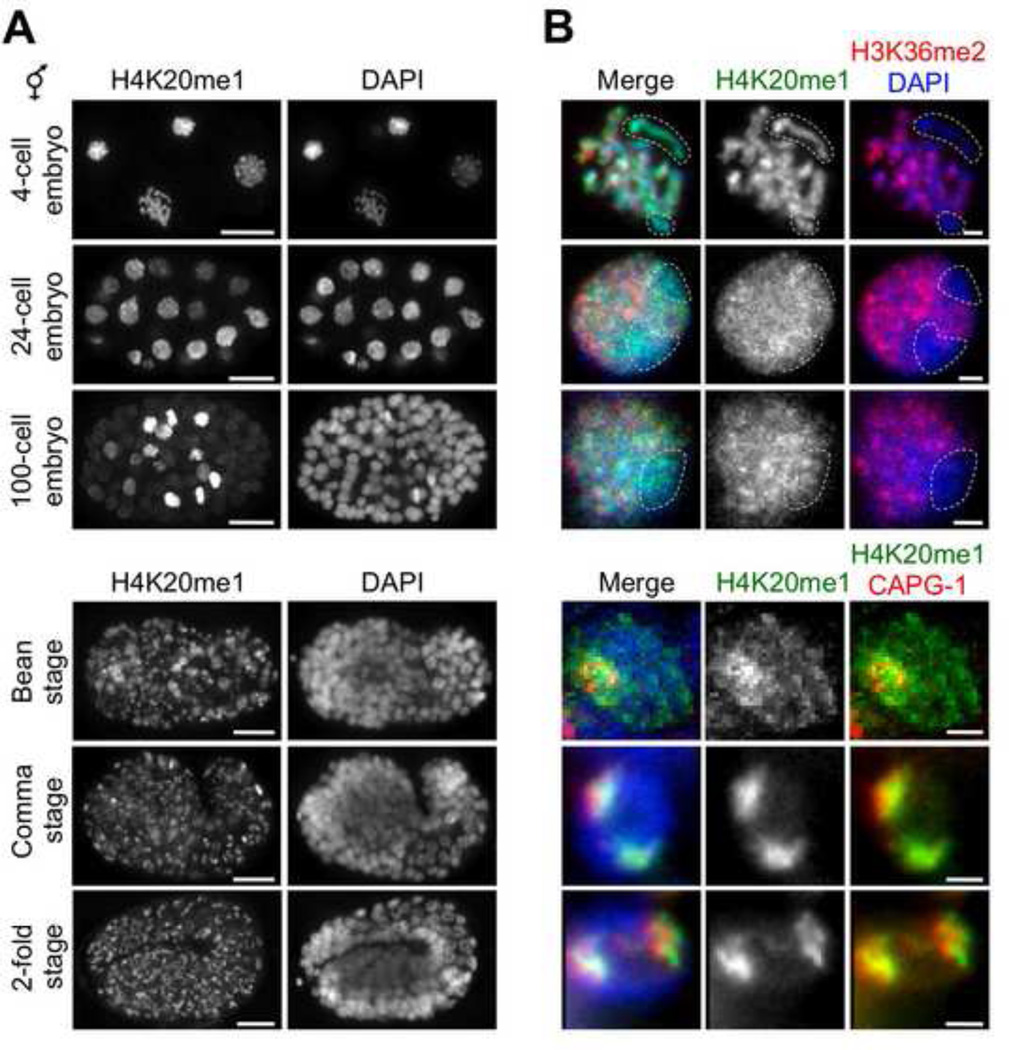
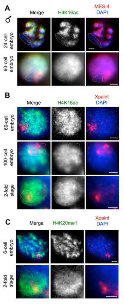
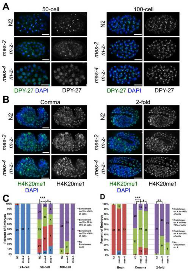
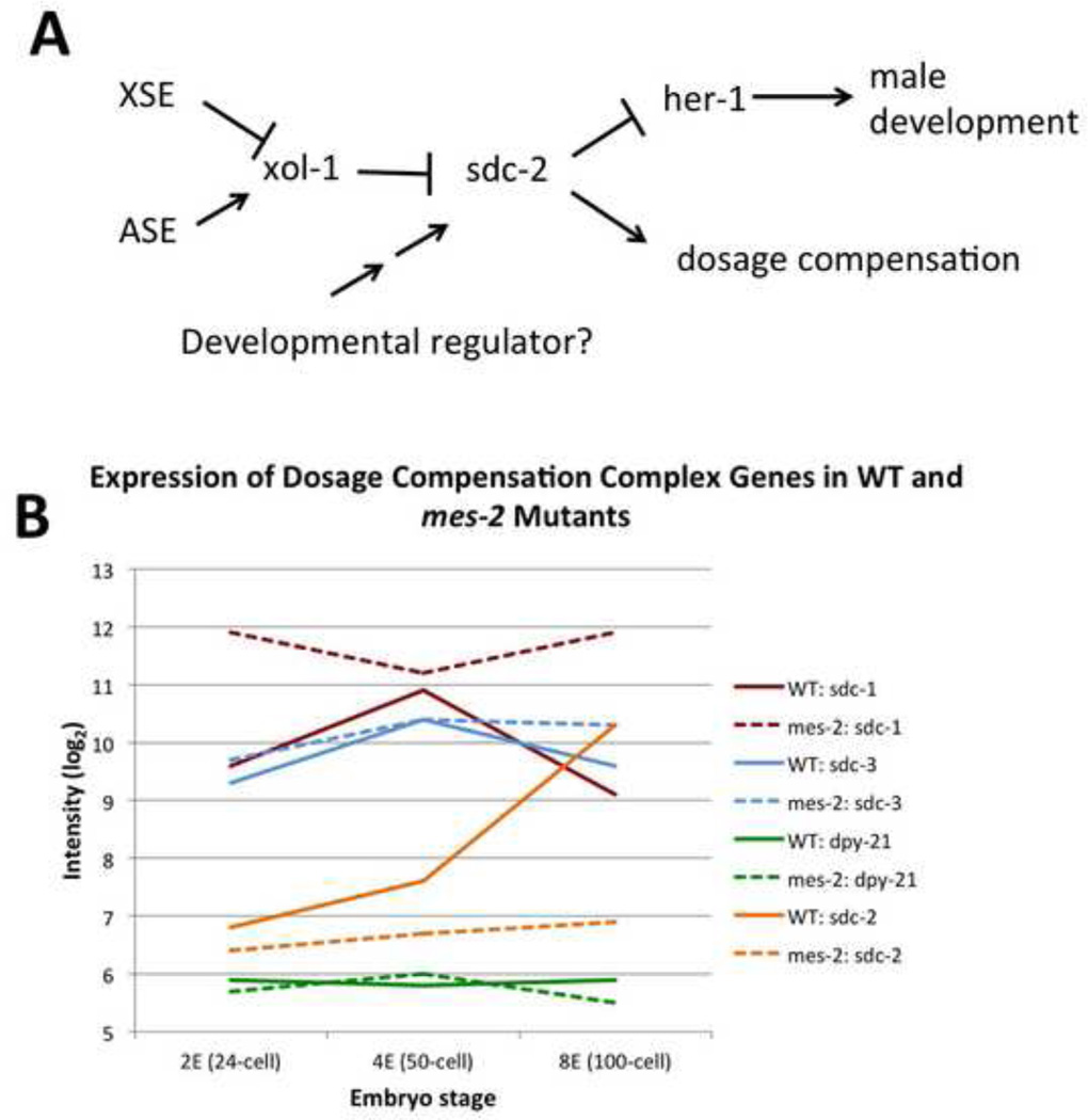
Dosage compensation (DC) equalizes X-linked gene expression between sexes. In Caenorhabditis elegans, the dosage compensation complex (DCC) localizes to both X chromosomes in hermaphrodites and downregulates gene expression 2-fold. The DCC first localizes to hermaphrodite X chromosomes at the 30-cell stage, coincident with a developmental transition from plasticity to differentiation. To test whether DC onset is linked to loss of developmental plasticity, we established a timeline for the accumulation of DC-mediated chromatin features on X (depletion of histone H4 lysine 16 acetylation (H4K16ac) and enrichment of H4K20 monomethylation (H4K20me1)) in both wild type and developmentally delayed embryos. Surprisingly, we found that H4K16ac is depleted from the X even before the 30-cell stage in a DCC-independent manner. This depletion requires the activities of MES-2, MES-3, and MES-6 (a complex similar to the Polycomb Repressive Complex 2), and MES-4. By contrast, H4K20me1 becomes enriched on X chromosomes several cell cycles after DCC localization to the X, suggesting that it is a late mark in DC. MES-2 also promotes differentiation, and mes-2 mutant embryos exhibit prolonged developmental plasticity. Consistent with the hypothesis that the onset of DC is linked to differentiation, DCC localization and H4K20me1 accumulation on the X chromosomes are delayed in mes mutant hermaphrodite embryos. Furthermore, the onset of hermaphrodite-specific transcription of sdc-2 (which triggers DC) is delayed in mes-2 mutants. We propose that as embryonic blastomeres lose their developmental plasticity, hermaphrodite X chromosomes transition from a MES protein-regulated state to DCC-mediated repression.

Keywords: Chromatin; Dosage compensation; Epigenetics; Pluripotency.


Figures

Figure 1
Figure 1. Acetylated H4K16 is depleted from the X chromosomes during all stages of hermaphrodite development
Antibody staining of H4K16ac (green) in wild type hermaphrodite embryos. The X chromosomes were identified by a lack of autosomal MES-4 (red, top panels) staining in young embryos or by the presence of the DCC component CAPG-1 (red, bottom panels) in older embryos. DAPI (blue) labels DNA. Grayscale images of H4K16ac and DAPI are shown for clarity. DIC images of staged embryos are shown (left column), and representative stained nuclei from each stage are shown. In all stages, less H4K16ac is present on the X chromosomes than the autosomes. For clarity, the X chromosomes are outlined with a dashed line in the merged and MES-4 images and indicated with an arrow in the grayscale H4K16ac images. Note that in the 2-cell embryo, one X chromosome (arrow) has reduced H4K16ac, while H4K16ac on the other X chromosome (arrowhead) resembles the autosomes. Scale bars equal 10 µm (whole embryo images) and 1 µm (nuclei).
Figure 2
Figure 2. H4K20me1 enrichment on the X chromosomes of hermaphrodite embryos occurs at the comma stage
Antibody staining of H4K20me1 (green), H3K36me2 (red) to label autosomes, the DCC component CAPG-1 (red) to label the X chromosomes, and DAPI (blue) to label DNA in wild type hermaphrodite embryos. (A) Projection images through the center z-slices of embryos stained with H4K20me1 and DAPI. (B) Representative stained nuclei from each stage shown in A. In 4- to 100-cell embryos, H4K20me1 is distributed evenly between the X chromosomes (outlined with dashed line) and autosomes. Variability in levels of H4K20me1 among nuclei in embryos younger than the bean stage is due to the cells being in different stages of the cell cycle. At the bean stage, H4K20me1 begins to be enriched on the X chromosomes in some nuclei. By the comma and 2-fold stages, H4K20me1 is enriched on the X chromosomes (labeled with CAPG-1) in most or all nuclei. Scale bars equal 10 µm (whole embryo images) and 1 µm (nuclei).
Figure 3
Figure 3. Histone H4 modifications on the male X chromosome
(A–B) H4K16ac (green) is depleted from the X chromosome (outlined with dashed line) in male him-8(e1489) embryos until the 60-cell stage. The X chromosome in male embryos was identified as the one chromosome per nucleus that did not have MES-4 (red) autosomal staining (A). After the 60-cell stage, H4K16ac levels on the male X are equivalent to autosomal levels (B). The embryos were identified as male by the presence of only one X territory, marked with Xpaint FISH, in every nucleus. (C) H4K20me1 levels are uniform between the X chromosome and autosomes in young male embryos. This modification does not become enriched on the male X chromosome at late morphogenesis stages. Scale bars equal 1 µm.
Figure 4
Figure 4. Prior to its localization to the X chromosomes, the DCC does not regulate H4K16ac levels
Antibody staining of H4K16ac (green), H3K36me2 (red) to label autosomes, and DAPI (blue) to label DNA in mutant hermaphrodite embryos. H4K16ac is reduced on the X chromosomes (outlined with a dashed line) compared to autosomes in 4- to 16-cell hermaphrodite embryos from wild type, sdc-1(y415) mutant, and capg-1, dpy-26, and dpy-28 RNAi-treated adults. Levels of H4K16ac are similar between the X chromosomes and autosomes in dpy-21(e428) mutant embryos. Scale bars equal 1 µm.
Figure 5
Figure 5. X chromosome H4K16ac levels are restored to autosomal levels in mes mutants
(A) Nuclei from 8- to 16-cell embryos stained with anti-H4K16ac (green). Xpaint FISH (red) labels the X chromosomes. In mes-2(bn11), mes-3(bn21), and mes-4(ok2326) M-Z− mutant and mes-6 RNAi-depleted hermaphrodite embryos, H4K16ac levels are equivalent between the X chromosomes (outlined with dashed line) and autosomes. (B) Immunofluorescent staining of H4K16ac (green) and CAPG-1 (X chromosomes, red). At the 2-fold stage, H4K16ac levels are depleted on the X chromosomes in mes-2(bn11) and mes-4(ok2326) M-Z− embryos and mes-3 and mes-6 RNAi-depleted embryos. Scale bars equal 1 µm.
Figure 6
Figure 6. The timing of DC onset is delayed in mes-2(bn11) and mes-4(ok2326) mutants
(A) At the 50-cell stage, the DCC component DPY-27 localizes to the X chromosomes in most cells in N2 embryos, but only a few cells in a mes-2(bn11) M-Z− and a mes-4(ok2316) M-Z− embryo. At the 100-cell stage, DPY-27 localizes to the X chromosomes in all cells in all three genotypes. (B) H4K20me1 becomes restricted to the X chromosomes in almost all nuclei by the comma stage in N2 embryos, but many nuclei do not have enrichment of H4K20me1 on the X chromosomes in comma stage and some 2-fold stage mes-2 M-Z− and mes-4 M-Z− embryos. (C) Quantification of DPY-27-stained N2, mes-2 M-Z−, and mes-4 M-Z− embryos. Each embryo was categorized based on the percent of its cells that demonstrated enrichment of DPY-27 on the X chromosomes. (D) Quantification of H4K20me1-stained N2, mes-2 M-Z−, and mes-4 M-Z-embryos. Scale bars equal 10 µm. Asterisks indicate level of statistical significance (one asterisk, P<0.05; two asterisks, P<0.005; three asterisks, P<0.001).
Figure 7
Figure 7. sdc-2 expression in mes-2(bn11) mutants
(A) Genetic pathway regulating SDC-2 expression and dosage compensation. (B) Analysis of gene expression of selected dosage compensation genes from a previously published dataset (Yuzyuk et al., 2009). Solid lines indicate wild type embryos and dashed lines indicate mes-2(bn11) mutant embryos.

Similar articles

Cited by

References

    1. Akhtar A, Becker PB. Activation of transcription through histone H4 acetylation by MOF, an acetyltransferase essential for dosage compensation in Drosophila. Mol Cell. 2000;5:367–375. - PubMed
    1. Arico JK, Katz DJ, van der Vlag J, Kelly WG. Epigenetic patterns maintained in early Caenorhabditis elegans embryos can be established by gene activity in the parental germ cells. PLoS Genet. 2011;7:el001391. - PMC - PubMed
    1. Barakat TS, Gribnau J. X chromosome inactivation in the cycle of life. Development. 2012;139:2085–2089. - PubMed
    1. Barakat TS, Gunhanlar N, Pardo CG, Achame EM, Ghazvini M, Boers R, Kenter A, Rentmeester E, Grootegoed JA, Gribnau J. RNF12 activates Xist and is essential for X chromosome inactivation. PLoS Genet. 2011;7:e1002001. - PMC - PubMed
    1. Baugh LR, Hill AA, Slonim DK, Brown EL, Hunter CP. Composition and dynamics of the Caenorhabditis elegans early embryonic transcriptome. Development. 2003;130:889–900. - PubMed

Publication types

LinkOut - more resources