Comparative Study
. 2014 Jan;20(1):144-53.
doi: 10.1097/01.MIB.0000436467.37566.48.

Anti-inflammatory effects of the Chinese herbal formula FAHF-2 in experimental and human IBD

Affiliations
Comparative Study

Anti-inflammatory effects of the Chinese herbal formula FAHF-2 in experimental and human IBD

Ying Song et al. Inflamm Bowel Dis. 2014 Jan.

Abstract

Background: Crohn's disease (CD) is a chronic inflammatory disease with increasing incidence in children. Current medications have potentially serious side effects, hence increasing interest in alternative therapies. We previously developed an herbal formula, FAHF-2, based on a classical traditional Chinese herbal formula Wu Mei Wan that has long been used in China to treat colitis. We investigated FAHF-2's potential anti-inflammatory effects.

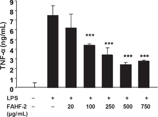
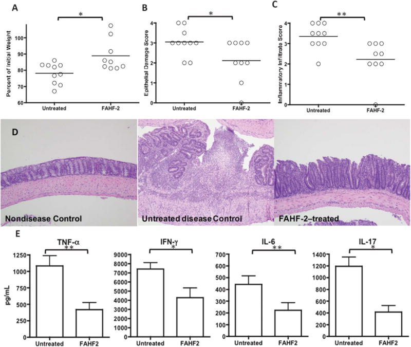
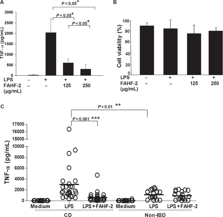
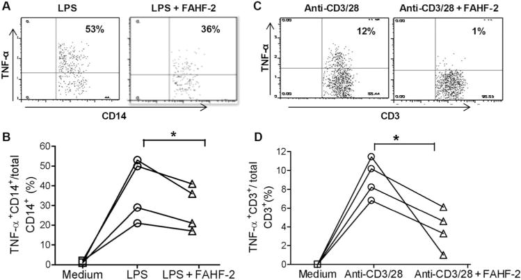
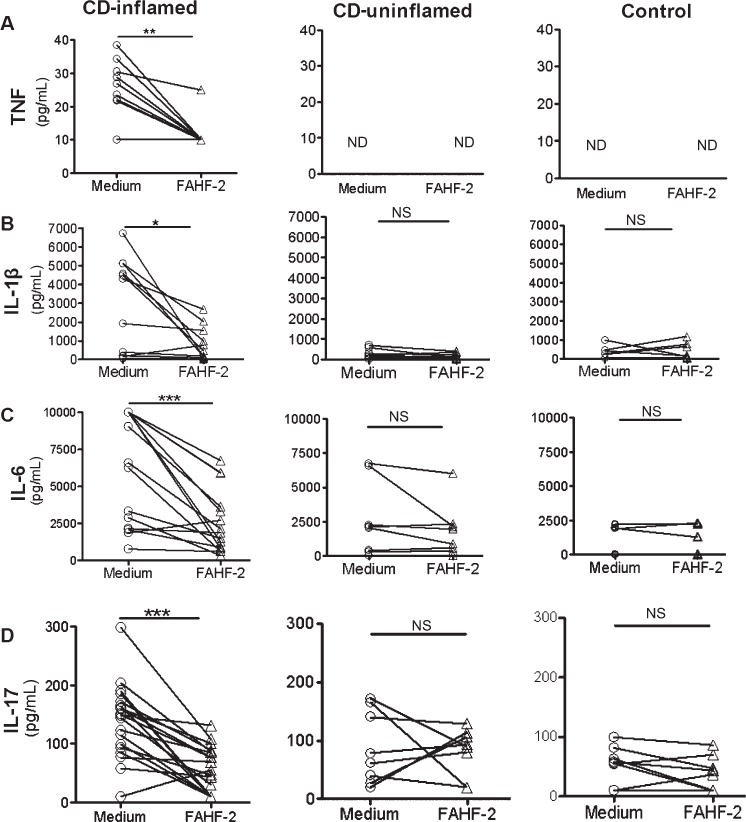
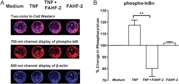
Methods: FAHF-2 efficacy was tested in vivo in the CD45RbRAG1 transfer colitis model. Weight loss, colonic histology, and cytokine production from mesenteric lymph nodes were assessed. Human peripheral blood mononuclear cells (PBMCs) and colonic biopsies were obtained from children newly diagnosed with CD and controls and cultured with or without FAHF-2. Cytokine levels were measured by multiplex immunoassay. The effect of FAHF-2 on TNF-α-producing cells was determined by flow cytometry. NF-κB signaling was investigated in human lamina propria mononuclear cells upon FAHF-2 treatment by In-Cell Western.

Results: FAHF-2-treated mice had decreased weight loss, improved histology, and reduced TNF-α, IL-17, IL-6, and IFN-γ production. In vitro treated PBMCs produced less TNF-α, IFN-γ, and IL-12. FAHF-2 reduced the TNF-α-producing monocytes and T cells. Inflamed CD biopsies produced less TNF-α, IL-17, IL-6, and IL-1β. These effects are because of decreased NF-κB activation.

Conclusions: FAHF-2 inhibited both adaptive and innate immune proinflammatory cytokine responses in PBMCs and inflamed CD mucosa due in part to blockage of NF-κB activation. FAHF-2 was effective in halting progression of colitis in a murine model. This study shows that FAHF-2 has potential as a novel treatment of CD.


Conflict of interest statement

X.-M. Li is a consultant for the FAI and has shares of US Patent PCT/US 05/08600 on FAHF-2 and Herbal Springs, LLC. The other authors have no conflicts of interest to disclose.

Figures

FIGURE 1
FIGURE 1
RAW264.7 cells were cultured with various doses of FAHF-2 (20–750 μg/mL) for 1 hour and then stimulated with 1 μg/mL of LPS for 24 hours. TNF-α levels were measured in the supernatants by enzyme-linked immunosorbent assay according to the manufacturer’s instructions (BD Bioscience). ***p< 0.001 vs. LPS alone.
FIGURE 2
FIGURE 2
The effects of FAHF-2 on weight loss, histology, and cytokine secretion from mesenteric lymph nodes in the CD45RBhi transfer colitis model. The final weight measurement as a percentage of initial weight (A), histological score for epithelial damage (B), and inflammatory infiltrate (C). Representative histological hematoxylin–eosin slides (D) at 10× from: nondiseased control (left): normal mucosa with no inflammatory infiltrate, untreated disease control (middle): mucosal ulceration with underlying mixed inflammatory infiltrate and disordered gland architecture, FAHF-2–treated (right): no ulceration, mixed inflammatory infiltrate, and architectural distortion improved from untreated disease control. MLN cytokine secretion in untreated and FAHF-2–treated mice with colitis: TNF-α, IFN-γ, IL-17, and IL-6 (E). *P< 0.05, ** P< 0.01.
FIGURE 3
FIGURE 3
Effect of FAHF-2 on TNF-α levels from PBMCs from CD subjects and controls. A. TNF-α levels as measured by enzyme-linked immunosorbent assay from 3 individual CD subject’s PBMCs cultured in medium alone, LPS alone, or LPS with FAHF-2 treatment (125 or 250 μg/mL). B. Cell viability after the cells were cultured. C. TNF-α levels from PBMCs from 26 CD and 18 controls. Symbols indicate individual subjects. Lines indicate the mean values for each condition. *P< 0.05, ** P< 0.01, *** P< 0.001.
FIGURE 4
FIGURE 4
Effect of FAHF-2 on TNF-α–producing cell populations measured by flow cytometry: changes in the ratio of TNF-α+CD14+/CD14+ cells with or without FAHF-2 treatment in an individual subject (A) and 4 subjects (B). Changes in the ratio of TNF-α+CD3+/CD3+ cells with or without FAHF-2 treatment in vitro in an individual subject (C) and 4 subjects (D). * P< 0.05.
FIGURE 5
FIGURE 5
Effect of FAHF-2 on cytokine levels from colonic biopsies from CD subjects and controls. Inflamed and noninflamed biopsies from CD and control subjects were cultured with or without 250 μg/mL of FAHF-2 for 24 hours. Cytokine levels were measured from the supernatants by cytometric bead array. Cytokine levels without (medium) and with treatment with FAHF-2 are shown for each cytokine: TNF-α, IL-1β, IL-6, and IL-17A. CD inflamed: n = 15; CD uninflamed: n = 8; non-IBD control (control): n = 9. ND, not detectable; NS, not significant. *P< 0.05, **P< 0.01, ***P< 0.001.
FIGURE 6
FIGURE 6
A. Representative In-Cell Western with 700 and 800 nm channels detecting phospho-IκB and β-actin, respectively, in LPMCs cultured in medium, stimulated with TNF, stimulated and treated with FAHF-2, or with FAHF-2 alone. B. Percentage of phosphorylated IκB measured by In-Cell Western after TNF stimulation and with FAHF-2 treatment as compared with medium alone (n = 3). All values normalized to β-actin. **P< 0.01.

Similar articles

Cited by

References

    1. Kappelman MD, Moore KR, Allen JK, et al. Recent trends in the prevalence of Crohn’s disease and ulcerative colitis in a commercially insured US population. Dig Dis Sci. 2013;58:519–525. - PMC - PubMed
    1. van der Zaag-Loonen HJ, Casparie M, Taminiau JA, et al. The incidence of pediatric inflammatory bowel disease in the Netherlands: 1999–2001. J Pediatr Gastroenterol Nutr. 2004;38:302–307. - PubMed
    1. Benchimol EI, Fortinsky KJ, Gozdyra P, et al. Epidemiology of pediatric inflammatory bowel disease: a systematic review of international trends. Inflamm Bowel Dis. 2011;17:423–439. - PubMed
    1. Benchimol EI, Guttmann A, Griffiths AM, et al. Increasing incidence of paediatric inflammatory bowel disease in Ontario, Canada: evidence from health administrative data. Gut. 2009;58:1490–1497. - PubMed
    1. Shih DQ, Targan SR, McGovern D. Recent advances in IBD pathogenesis: genetics and immunobiology. Curr Gastroenterol Rep. 2008;10:568–575. - PMC - PubMed

Publication types

MeSH terms