. 2014 Jan 3;289(1):540-51.
doi: 10.1074/jbc.M113.518795. Epub 2013 Nov 19.

Stabilization of the dimeric birch pollen allergen Bet v 1 impacts its immunological properties

Affiliations

Stabilization of the dimeric birch pollen allergen Bet v 1 impacts its immunological properties

Stefan Kofler et al. J Biol Chem. .

Abstract

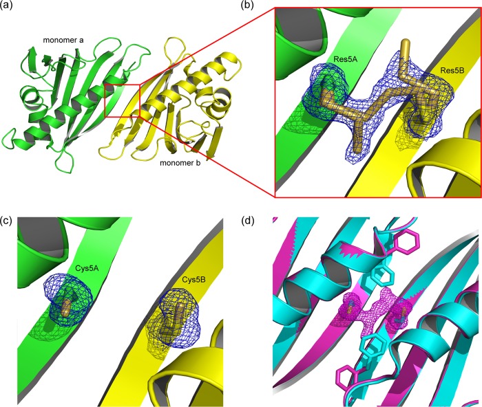
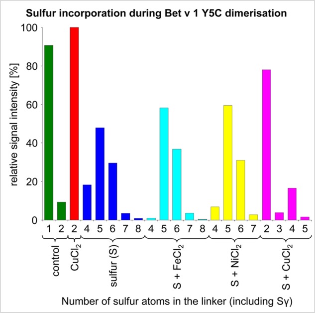
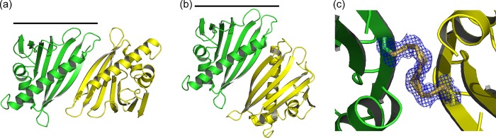
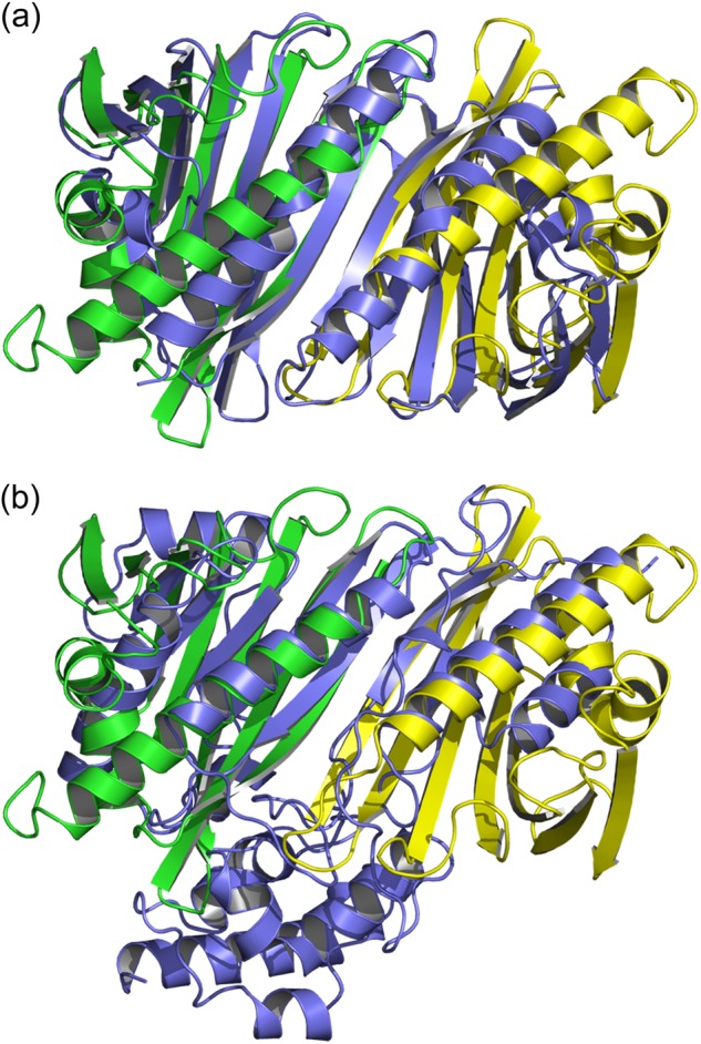
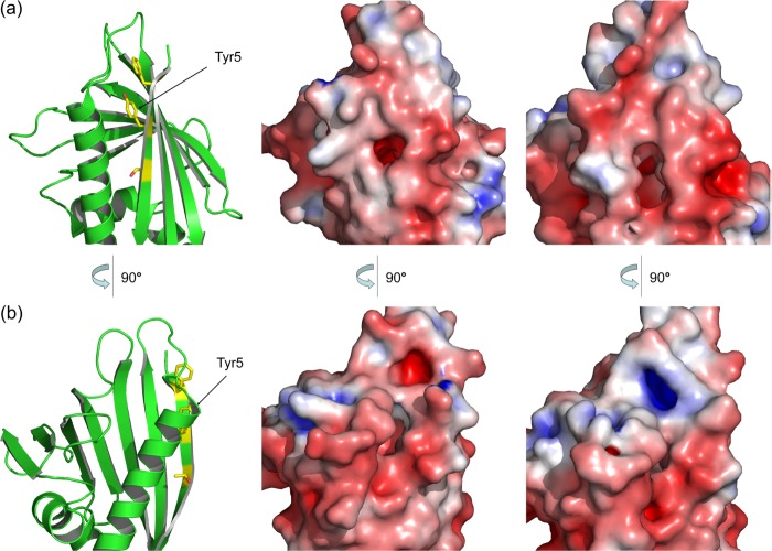
Many allergens share several biophysical characteristics, including the capability to undergo oligomerization. The dimerization mechanism in Bet v 1 and its allergenic properties are so far poorly understood. Here, we report crystal structures of dimeric Bet v 1, revealing a noncanonical incorporation of cysteine at position 5 instead of genetically encoded tyrosine. Cysteine polysulfide bridging stabilized different dimeric assemblies, depending on the polysulfide linker length. These dimers represent quaternary arrangements that are frequently observed in related proteins, reflecting their prevalence in unmodified Bet v 1. These conclusions were corroborated by characteristic immunologic properties of monomeric and dimeric allergen variants. Hereby, residue 5 could be identified as an allergenic hot spot in Bet v 1. The presented results refine fundamental principles in protein chemistry and emphasize the importance of protein modifications in understanding the molecular basis of allergenicity.

Keywords: Allergen; Crystal Structure; Dimerization; Mass Spectrometry (MS); Noncanonical Amino Acid Incorporation; Polysulfide Linking; Position-specific Alteration of Genetic Code; Post-translational Modification; Protein Assembly.


Figures

FIGURE 1.
FIGURE 1.
Identification of dimeric Bet v 1 during WT protein preparation. a, separation of monomeric and dimeric Bet v 1 via ion exchange (top panel, peak corresponding to dimer indicated by black lines) and size exclusion chromatography (bottom panel). b, analysis of size exclusion chromatography fractions via native (top panel) and SDS (middle and bottom panels) PAGE. Comparison of reducing (middle panel) and nonreducing (bottom panel) SDS-PAGE demonstrates the reduction sensitivity of the dimer. c, SDS- and clear native-PAGE revealed stability of the correctly folded dimer under reducing conditions (bottom panel, right side). The bands corresponding to dimeric Bet v 1 are boxed. Lane M, marker; lanes 17 and 35, molecular mass of the marker band in kDa.
FIGURE 2.
FIGURE 2.
Tetrasulfide dimer interface in the absence and presence of reducing agent. a, cartoon representation of the assembly in the tetrasulfide dimer. The β1-strands of the monomers (green and yellow) elongate the antiparallel β-sheet over the whole quaternary structure. b, close-up view of the dimer interface. The tetrasulfide bridge exhibits partial breaks between SγA-SδA and SδB-SγB. c, the dimer interface after reduction, with the linker electron density vanished. The residual electron density at residue 5 fits cysteines. d, comparison of Phe3 rotamers in the intact (magenta) and reduced (cyan) tetrasulfide dimer. Reduction leads to an approximately 120° flip of Phe3 toward residue 5. The presence of the linker (indicated by its density) keeps Phe3 in the same conformation as observed in the WT structure.
FIGURE 3.
FIGURE 3.
Variation of sulfur atom incorporations in Bet v 1 Y5C. The length of the linkers is dependent on the dimerization protocol. CuCl2 favors disulfide formation, reducing the amount of incorporated sulfur. By contrast, the addition of NiCl2 and FeCl2 had only a little influence on the polysulfide pattern.
FIGURE 4.
FIGURE 4.
Comparison of the quaternary arrangement in tetra- and nonasulfide bridged Bet v 1. a, the tetrasulfide bridged dimer is stabilized by anti-parallel β-sheet formation, with an interface of 536 Å2. b, the nonasulfide bridged Bet v 1 is rotated along the sulfide bridge for ∼135°, resulting in a larger interface (670 Å2) and the loss of the intermolecular β-sheet extension. c, all nine sulfur atoms are resolved in the 1.17 Å resolution electron density.
FIGURE 5.
FIGURE 5.
Structural homologues with folds equivalent to the tetrasulfide dimer. Bet v 1 is shown in green and yellow, and the homologues are in slate blue. a, SMU.440 from S. mutans forms stable homodimers in solution. b, the single chain arginine kinase from L. polyphemus exhibits an antiparallel β-sheet capped by two long helices, similar to the elongated sheet in the tetrasulfide dimer.
FIGURE 6.
FIGURE 6.
Identical ligand binding capacity of monomeric and dimeric Bet v 1. Changes in the fluorescence signal (ΔF/F0) of ANS induced by the presence of DXC in different molar ratios. Titration of both the monomer (circles) and the dimer (squares) gave comparable results for both ANS binding (ratio 1:0) as well as ANS replacement.
FIGURE 7.
FIGURE 7.
Accessibility of IgE epitopes in the tetrasulfide dimer. Residues corresponding to IgE epitopes are highlighted as sticks and surface. a, b, and d, three of four reported epitopes allow cross-linking of univalent IgE antibodies via dimeric Bet v 1 (shown as red surface) (38, 40). c, by contrast, a fourth epitope at the C-terminal α-helix of Bet v 1 (blue surface) is partly buried by the dimer interface, restricting simultaneous binding of two IgE (39). d, a schematic model for IgE cross-linking facilitated by dimeric Bet v 1 on the surface of an effector cell, mediated by FcϵRI, based on Ref. .
FIGURE 8.
FIGURE 8.
IgE binding to monomeric and dimeric Bet v 1a variants tested via ELISA and rat basophile leukemia mediator release assay. a, ELISA. The amount of protein needed for half-maximal antibody binding is plotted. Reduction of IgE reactivity correlated with alteration of the surface patch near residue 5. The strongest effect was obtained with Y5Cglu. b, rat basophile leukemia (RBL) mediator release assay. The amount of protein that is needed for half-maximal mediator release (expressed as a percentage of total enzyme content in the cells) is plotted. No significant differences between monomeric and dimeric Bet v 1a variants could be detected. Y5Cglu induced a significant decrease in mediator release. Bet a, WT Bet v 1a; Y5Cred, monomeric Bet v 1a Y5C (reduced); dimer, dimeric protein obtained from WT Bet v 1a preparation; Y5C, dimeric Bet v 1a obtained from mutant preparation; Y5Cglu, Bet v 1a Y5C with Cys5 oxidized, and capped, with glutathione.
FIGURE 9.
FIGURE 9.
Surface changes induced by the Y5C mutation. The Y5C exchange propagates to the conformation of neighboring amino acids (cf. Fig. 2d), generating pronounced changes in shape and charge distribution at its environment. a and b, comparison of Bet v 1a and Bet v 1a Y5C. Two views of the affected region are shown. Left, schematic representation of Bet v 1a for orientation. Middle, Bet v 1a. Right, Bet v 1a Y5C.
FIGURE 10.
FIGURE 10.
Different Bet v 1 variants induce distinct cytokine releases in primary dendritic cells. Cytokine secretion after stimulation of human primary DCs with 50 μg/ml of native Bet v 1a, Bet Y5F, or Bet A5C is shown. Uninduced cells (control) as well as cells stimulated with 10 ng/ml LPS and 30 ng/ml thymic stromal lymphopoietin were included as controls. Single values (squares) and the mean (bars) are depicted. Statistical significance was calculated with the Student's t test for each comparison of Bet v 1 versus Bet Y5F and Bet Y5C and for Bet Y5F versus Bet Y5C (*, p < 0.05; **, p < 0.01; ***, p < 0.005). a, TH1 priming cytokines IL-12 and IL-6. b, cytokines TFN-α and MCP-1, inducing a mixed polarization. c, TH2 priming cytokines MDC and TARC.

Similar articles

Cited by

References

    1. Schein C. H., Ivanciuc O., Midoro-Horiuti T., Goldblum R. M., Braun W. (2010) An Allergen Portrait Gallery. Representative structures and an overview of IgE binding surfaces. Bioinform. Biol. Insights 4, 113–125 - PMC - PubMed
    1. Shakib F., Ghaemmaghami A. M., Sewell H. F. (2008) The molecular basis of allergenicity. Trends Immunol. 29, 633–642 - PubMed
    1. Sokol C. L., Barton G. M., Farr A. G., Medzhitov R. (2008) A mechanism for the initiation of allergen-induced T helper type 2 responses. Nat. Immunol. 9, 310–318 - PMC - PubMed
    1. Verdino P., Westritschnig K., Valenta R., Keller W. (2002) The cross-reactive calcium-binding pollen allergen, Phl p 7, reveals a novel dimer assembly. EMBO J. 21, 5007–5016 - PMC - PubMed
    1. Lascombe M. B., Grégoire C., Poncet P., Tavares G. A., Rosinski-Chupin I., Rabillon J., Goubran-Botros H., Mazié J. C., David B., Alzari P. M. (2000) Crystal structure of the allergen Equ c 1. A dimeric lipocalin with restricted IgE-reactive epitopes. J. Biol. Chem. 275, 21572–21577 - PubMed

Publication types