. 2014 Jan 3;289(1):565-80.
doi: 10.1074/jbc.M113.466326. Epub 2013 Nov 19.

Mutational and structural analysis of the tandem zinc finger domain of tristetraprolin

Affiliations

Mutational and structural analysis of the tandem zinc finger domain of tristetraprolin

Wi S Lai et al. J Biol Chem. .

Abstract

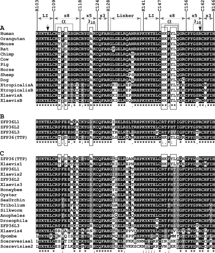
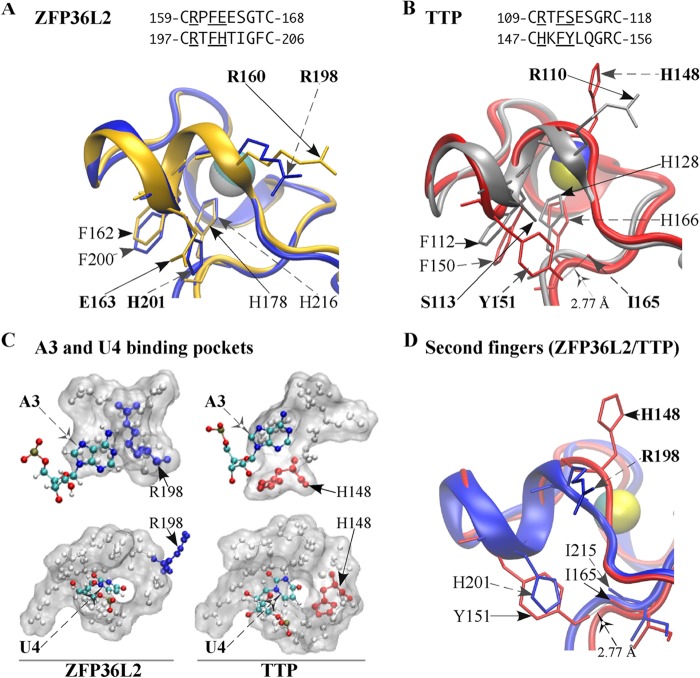
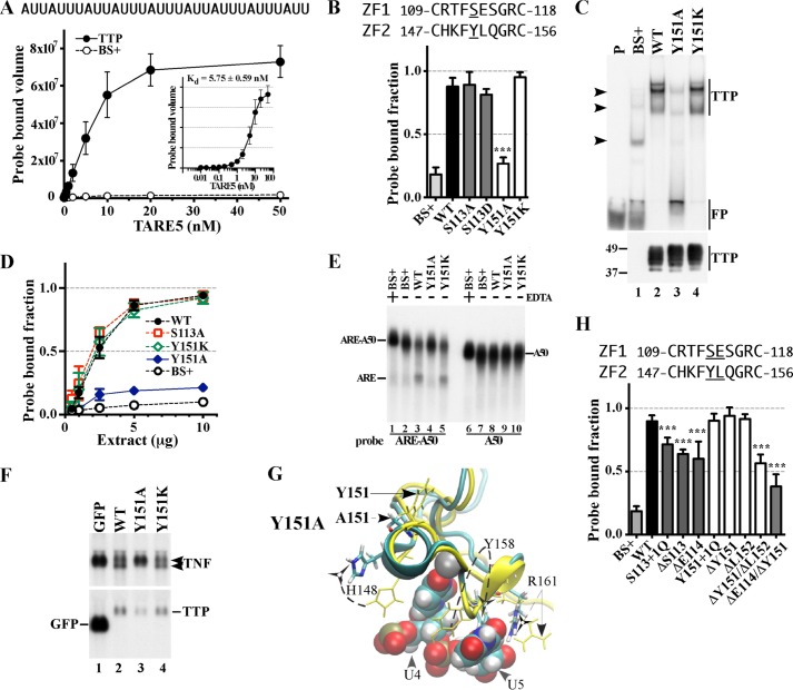
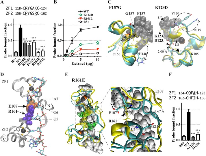
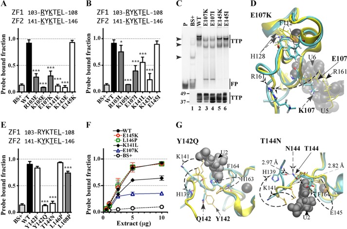
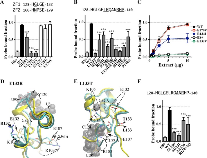
Tristetraprolin (TTP), the best known member of a class of tandem (R/K)YKTELCX8CX5CX3H zinc finger proteins, can destabilize target mRNAs by first binding to AU-rich elements (AREs) in their 3'-untranslated regions (UTRs) and subsequently promoting deadenylation and ultimate destruction of those mRNAs. This study sought to determine the roles of selected amino acids in the RNA binding domain, known as the tandem zinc finger (TZF) domain, in the ability of the full-length protein to bind to AREs within the tumor necrosis factor α (TNF) mRNA 3'-UTR. Within the CX8C region of the TZF domain, mutation of some of the residues specific to TTP, not found in other members of the TTP protein family, resulted in decreased binding to RNA as well as inhibited mRNA deadenylation and decay. Evaluation of simulation solution models revealed a distinct structure in the second zinc finger of TTP that was induced by the presence of these TTP-specific residues. In addition, mutations within the lead-in sequences preceding the first C of highly conserved residues within the CX5C or CX3H regions or within the linker region between the two fingers also perturbed both RNA binding and the simulation model of the TZF domain in complex with RNA. We conclude that, although the majority of conserved residues within the TZF domain of TTP are required for productive binding, not all residues at sequence-equivalent positions in the two zinc fingers of the TZF domain of TTP are functionally equivalent.

Keywords: AU-rich Elements; Cytokine; Inflammation; RNA Turnover; RNA-Protein Interaction; RNA-binding Protein; Tumor Necrosis Factor (TNF).


Figures

FIGURE 1.
FIGURE 1.
Alignments of the tandem CCCH zinc fingers of TTP family members. A, an alignment of TZF domains from vertebrate TTP orthologues that were readily available using RefSeq proteins and expressed sequence tags. The numbers above the human TTP TZF sequence represent the residue position in NP_003398.1, and only the first residues of the lead-in sequence to each finger and the CCCH residues are indicated. The following are also indicated: the lead-in (LI) sequences to each finger; the interval distances within the CCCH motifs and the linker sequence; and the locations of those residues that form the helices (29). Note that in TTP the α-helix of finger 2 is indicated by the hatched lines. The two downward arrows indicate residues Glu107 and Arg161, whose ionic interaction stabilizes the two fingers (see Fig. 4B). The seven residues specific to TTP are highlighted in white background boxes. B, an alignment of the TZF domains of the four TTP family members known to exist in mice. C, an alignment of the TZF domains of TTP family members that occur in various more distant eukaryotes. The TTP family members from mouse and X. laevis were used as representatives of vertebrate family members; a sampling of yeasts, insects, and aquatic invertebrates was added. Alignments were generated using ClustalW and BoxShade. Asterisks, amino acid identity at a site; colons, a high degree of chemical conservation at that site; single dots, a lower degree of chemical conservation at that site. The GenBankTM accession numbers for sequences used in this figure are as follows: for TTP, human (NP_003398.1), orangutan (XP_002834549.2), mouse (NP_035886), rat (NP_579824), chimpanzee (XP_001136016), cow (NP_776918), pig (NP_001161891), horse (translation of CD536573.1), sheep (NP_001009765), dog (XP_541624), Silurana tropicalis (NP_001106542), X. laevis (NP_001081884); for other mouse TTP family members, ZFP36L1 (NP_031590), ZFP36L2 (NP_001001806), ZFP36L3 (NP_001009549); for other X. laevis TTP family members, ZFP36L1 (NP_001084214), ZFP36L2 (NP_001080610), C3H-4 (NP_001081889); for other organisms, oyster unnamed protein (translation of AAB69448.1), sea urchin unnamed protein (XP_782811.1), Schizosaccharomyces pombe Zfs1p (NP_596453), CTH1 and CTH2 from S. cerevisiae (AAB39897 and AAB39898, respectively); insect family members that are orthologues of the Drosophila melanogaster protein Tis11 (NP_511141), Apis mellifera (NP_001121248.1), Tribolium castaneum (XP_968440.1), Bombyx mori (translation of AK382012.1), Anopheles gambiae (XP_309752.3).
FIGURE 2.
FIGURE 2.
Backbone superposition of TZF structural models of human ZFP36L2 (TIS11d) and TTP. The models shown were based on the initial RNA-bound structure of the TZF domain of ZFP36L2 (see “Experimental Procedures”). In A and B, finger 1 side chains are indicated by solid arrows, finger 2 side chains are indicated by dashed arrows, and zinc atoms are shown as spheres. A, superposition of the two fingers of ZFP36L2 (Arg153–Glu220). The ribbon diagram of the peptide backbone and selected side chains of finger 1 are depicted in gold, and those of finger 2 are in blue. Finger 1 side chains of Arg160 and Glu163, finger 2 side chains of Arg198 and His201, the zinc-coordinating residues His178/His216, and their respective stacking interacting residues Phe162/Phe200, are shown for both fingers. B, superposition of the two fingers of TTP (Arg103–Glu170). The ribbon diagram of the peptide backbone and selected side chains of finger 1 are depicted in silver, and those of finger 2 are in red. Finger 1 side chains of Arg110 and Ser113, finger 2 side chains of His148 and Tyr151, backbone of Ile165, the zinc-coordinating residues His128/His166, and their respective stacking interacting residues Phe112/Phe150 are shown for both fingers. The hydrogen bond formed by the side chain of Tyr151, and the backbone of Ile165 is also illustrated. C, surface representation of the binding pockets for the ARE bases A3 and U4. Nucleosides A3 and U4 are shown in colors. The side chain of Arg198 in finger 2 of ZFP36L2 is blue, and the side chain of His148 in finger 2 of TTP is red. Residues within the TZF domain of TTP or ZFP36L2 forming these binding pockets are listed in Table 1. D, Superposition of the second fingers of ZFP36L2 and TTP. The ribbon diagram of the peptide backbone and selected side chains of TTP finger 2 are shown in red, and those of ZFP36L2 are in blue. The hydrogen bond formed by the side chain of Tyr151 and the backbone of Ile165 is shown. Side chains of TTP shown are indicated by solid arrows, and side chains of ZFP36L2 are shown by dashed arrows. Zinc is displayed as spheres.
FIGURE 3.
FIGURE 3.
Effect of mutated residues in the CX8C regions of the human TTP TZF domain on RNA binding and RNA stability. A, equilibrium binding of probe TARE5 (0.01–50 nm) to extracts of HEK293 cells transfected with TTP or vector (BS+) as described under “Experimental Procedures.” The sequence of the RNA probe is depicted above the graph. The inset shows the TTP probe binding curve, in which the x axis was transformed into antilog, and the apparent dissociation constant (Kd ± S.D.) was calculated from quantification of three independent experiments. B, the binding of protein to RNA is expressed as the probe-bound fraction. Above the graph are the amino acid sequences of the intervals. The underlined residues are those that were mutated and whose effects on binding are shown in the bar graph. The results from mutants of finger 1 are shown as gray columns, and those of finger 2 are shown as white columns. The results (mean ± S.D. (error bars)) are from three similar gel shift assays using 0.2 nm probe TARE5. C, the top panel shows representative gel shift assays using extracts from HEK293 cells transfected with vector alone (BS+), WT human TTP, or various mutant TTP constructs. Cytosolic extracts were incubated with 0.2 nm probe TARE5 as described under “Experimental Procedures” before loading on a non-denaturing acrylamide gel. A sample that contained probe alone in buffer was also loaded (lane P). The migration positions of the TTP-ARE complex (TTP) and the free probe (FP) are indicated to the right with vertical bars, and the three major endogenous protein-ARE complexes are labeled with arrowheads to the left. The bottom panel shows the relative amount of WT TTP protein and its mutants used in the binding reaction, as determined by Western blotting. D, binding assays with 0.2 nm TARE5 probe in serially diluted extract, where total protein ranges from 10 to 1 μg, were performed as described under “Experimental Procedures.” The probe-bound fractions (mean ± S.D.) are from three similar gel shift assays. E, shown is a deadenylation assay using these extracts (5 μg of protein/sample). In lanes 1 and 6, 20 mm EDTA was present during the incubation to inhibit the deadenylase activity. In lanes 1–5, the ARE-A50 probe was used. The full-length probe ARE-A50 and its deadenylated product ARE are indicated to the left. Probe A50 was used in the samples shown in lanes 6–10, and the migration of the probe is indicated to the right. F, Northern blots of a HEK293 cell co-transfection of CMV.TNF with control plasmid GFP (lane 1), WT TTP (lane 2), Y151A (lane 3), or Y151K (lane 4). Total cellular RNA was prepared from the cells, and 10 μg of RNA was used for each lane. The blots were hybridized with a TNF cDNA probe (top) or a mouse TTP cDNA probe and a GFP cDNA probe together (bottom). The migration positions of the TNF mRNA and the TTP mRNA are indicated to the right, and that of the GFP RNA is shown to the left. G, superposition of WT TTP finger 2 and its mutant Y151A structure ensembles. The wild-type TTP peptide backbone ribbon and side chains are in yellow, and the mutant Y151A peptide backbone ribbon is in cyan, with side chains in colors. Zinc atoms (superimposed) are shown as silver spheres. Also shown are ARE nucleosides U4 and U5 (RMSD value calculated from superposition of the TZF domains of Y151A with RNA-bound WT TTP is 8.98 Å). H, the binding of protein to RNA is expressed as the probe-bound fraction. The results (mean ± S.D.) are from three similar gel shift assays. The results from mutants of finger 1 are shown as gray columns, and those of finger 2 are shown as white columns. Other details are similar to those described in B.
FIGURE 4.
FIGURE 4.
Effects of substitution mutants in the CX5CX3H intervals of the TTP TZF domain on RNA binding. The binding of protein to RNA is expressed as the probe-bound fraction. Above the graphs are the amino acid sequences of the intervals under study. The underlined residues are those that were mutated and whose effects on binding are shown in the bar graphs. The results from mutants of finger 1 are shown as gray columns, and those of finger 2 are shown as white columns. A, substitution mutants in the CX5C intervals. The results (mean ± S.D. (error bars)) are from four similar gel shift assays using 0.2 nm probe TARE5. B, binding assays with 0.2 nm TARE5 probe in serially diluted extracts, with total protein ranging from 10 to 1 μg. The probe-bound fractions (mean ± S.D.) are from four similar gel shift assays. C, superposition of the structural ensembles of finger 2 of WT TTP and mutant P157G (the RMSD is 10.37 Å; RMSD values were calculated from the superposition of the TZF domains of each mutant TTP with RNA-bound WT TTP) or finger 1 of WT TTP and mutant K123D (RMSD is 2.86 Å). The ribbon diagram of the WT TTP peptide backbone and side chains (sticks) are in yellow, the ribbon diagram of the mutant peptide backbone is in cyan, and side chains are shown in colors. RNA nucleosides are shown as gray spheres. Zinc atoms are shown as silver and copper (in P157G) or black and copper (in K123D) spheres. D, ribbon diagram of the peptide backbone of the human TTP TZF domain in complex with the ARE nonamer (sticks). The side chains of Glu107 (orange spheres, at the lead-in to finger 1) and Arg161 (purple spheres, at the C+5 position of the finger 2 CX5C region) are shown. Dashed arrows indicate nucleosides U5, U6, and A7 that interact with Glu107 and Arg161. Zinc atoms (black spheres) and the zinc-coordinating residues (ball and stick) of each finger are also displayed. E, superposition of the peptide backbone of the TZF domains of WT TTP (yellow ribbon) in complex with the ARE nonamer (surface representation) and the mutant R161E (cyan ribbon; RMSD is 4.66 Å). Zinc atoms are shown as black and copper spheres. Residues Glu107 and Arg161 (yellow mesh in the WT TZF domain) and Glu107 and Glu161 (green mesh in the mutant R161E) are shown (left). The right panel depicts the detailed view of the interaction between Arg161 and Glu107 (yellow sticks) in the WT and the non-interacting Glu107 and Glu161 (color sticks) in the mutant R161E. RNA nucleosides U4, U5, U6, and A7 (gray spheres) are indicated. F, substitution mutants in the CX3H intervals. The results (mean ± S.D.) are from four similar gel shift assays.
FIGURE 5.
FIGURE 5.
Effects of substitution mutants in the lead-in sequences to the zinc fingers on RNA binding. The binding of protein to RNA is expressed as the probe-bound fraction. Above the graphs are the amino acid sequences of the lead-in amino acids. The underlined residues are those that were mutated and whose effects on binding are shown in the bar graphs. The results from mutants of finger 1 are shown as gray columns, and those of finger 2 are shown as white columns. A and B, effects of mutant and WT TTP proteins, expressed as the probe-bound fraction. A, effects of charge reversal mutants; B, effects of neutral mutants. The results (mean ± S.D. (error bars)) are from three similar gel shift assays. C, the top panel shows representative gel shift assays using extracts from HEK293 cells transfected with vector alone (BS+), WT human TTP, or various mutant TTP constructs. Cytosolic extracts were incubated with 0.2 nm probe TARE5 as described under “Experimental Procedures” before loading on a non-denaturing acrylamide gel. A sample of probe alone in buffer was also loaded (lane P). Migration of the TTP-ARE complex (TTP) and the free probe (FP) are indicated on the right by vertical bars, and the three major endogenous protein-ARE complexes are labeled with arrowheads on the left. The bottom panel shows the relative amount of WT TTP protein and its mutants used in the binding reaction, as determined by Western blotting. D, superposition of WT TTP finger 1 and mutant E107K finger 1 structural ensembles (RMSD is 6.24 Å; RMSD values were calculated from superposition of the TZF domains of each mutant TTP with RNA-bound WT TTP). Wild-type TTP peptide backbone ribbon and side chains are in yellow, mutant E107K peptide backbone ribbon is in cyan, and side chains are in colors. RNA nucleosides U5 and U6 are shown as gray spheres. Zinc atoms are shown as black and copper spheres. E, comparisons in RNA binding are shown between mutant and WT TTP proteins; the results (mean ± S.D.) are from three similar gel shift assays. F, binding assays with 0.2 nm TARE5 probe in serially diluted extract with 1–10 μg of total protein. The probe-bound fractions (mean ± S.D.) are from three similar gel shift assays. G, superposition of WT TTP finger 2 and the Y142Q (RMSD value is 7.41 Å) or T144N (RMSD value is 10.66 Å) mutant model peptides. Color schemes are the same as in C, and RNA nucleoside U2 is shown. Zinc atoms are shown as silver and copper spheres.
FIGURE 6.
FIGURE 6.
Effect of mutated residues or altered distances in the interfinger linker region of the TTP TZF domain on RNA binding. Above the graphs are the amino acid sequences of the intervals. The underlined residues are those that were mutated and whose effects on binding are shown in the bar graphs. A, comparisons between substitution mutants in the four-residue stretch after the CCCH motifs with WT TTP protein, expressed as probe-bound fraction. The results (mean ± S.D. (error bars)) are from three similar gel shift assays. The results from mutants of residues immediately after finger 1 are shown as gray columns, and results from those after finger 2 are shown as white columns. B, comparisons between substitution mutants in the linker region with WT TTP protein, expressed as probe-bound fraction. The results (mean ± S.D.) are from three similar gel shift assays. C, binding assays with 0.2 nm TARE5 probe in serially diluted extract with 1–10 μg of total protein. The probe-bound fractions (mean ± S.D.) are from three similar gel shift assays. Shown is superposition of the models of finger 1 of WT TTP and mutant peptides E132R (in D; RMSD value for the TZF domains from superposition with RNA-bound WT TTP is 5.27 Å) or L133T (in E; RMSD value is 6.93 Å). The WT TTP peptide backbone (ribbon) and side chains (sticks) are yellow, the mutant peptide backbone is cyan, and the side chains are in colors. Zinc atoms are shown as silver and copper spheres. RNA nucleosides (U9 and U8 in D; U6 and U5 in E) are shown as gray spheres. F, comparisons between deletion or addition mutants in the linker region with WT TTP protein, expressed as probe-bound fraction. The results (mean ± S.D.) are from three similar gel shift assays.

Similar articles

Cited by

References

    1. Lai W. S., Stumpo D. J., Blackshear P. J. (1990) Rapid insulin-stimulated accumulation of an mRNA encoding a proline-rich protein. J. Biol. Chem. 265, 16556–16563 - PubMed
    1. Taylor G. A., Lai W. S., Oakey R. J., Seldin M. F., Shows T. B., Eddy R. L., Jr., Blackshear P. J. (1991) The human TTP protein. Sequence, alignment with related proteins, and chromosomal localization of the mouse and human genes. Nucleic Acids Res. 19, 3454. - PMC - PubMed
    1. Heximer S. P., Forsdyke D. R. (1993) A human putative lymphocyte G0/G1 switch gene homologous to a rodent gene encoding a zinc-binding potential transcription factor. DNA Cell Biol. 12, 73–88 - PubMed
    1. Ma Q., Herschman H. R. (1991) A corrected sequence for the predicted protein from the mitogen-inducible TIS11 primary response gene. Oncogene 6, 1277–1278 - PubMed
    1. DuBois R. N., McLane M. W., Ryder K., Lau L. F., Nathans D. (1990) A growth factor-inducible nuclear protein with a novel cysteine/histidine repetitive sequence. J. Biol. Chem. 265, 19185–19191 - PubMed

Publication types

MeSH terms

LinkOut - more resources