. 2014 Jan 3;289(1):379-86.
doi: 10.1074/jbc.M113.523209. Epub 2013 Nov 19.

The DNA methyltransferase Dnmt1 directly interacts with the SET and RING finger-associated (SRA) domain of the multifunctional protein Uhrf1 to facilitate accession of the catalytic center to hemi-methylated DNA

Affiliations

The DNA methyltransferase Dnmt1 directly interacts with the SET and RING finger-associated (SRA) domain of the multifunctional protein Uhrf1 to facilitate accession of the catalytic center to hemi-methylated DNA

Ahmet Can Berkyurek et al. J Biol Chem. .

Abstract

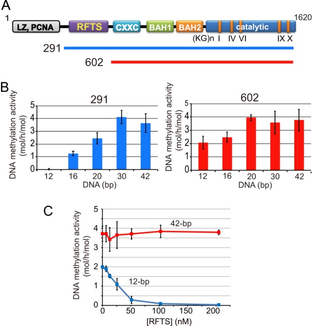
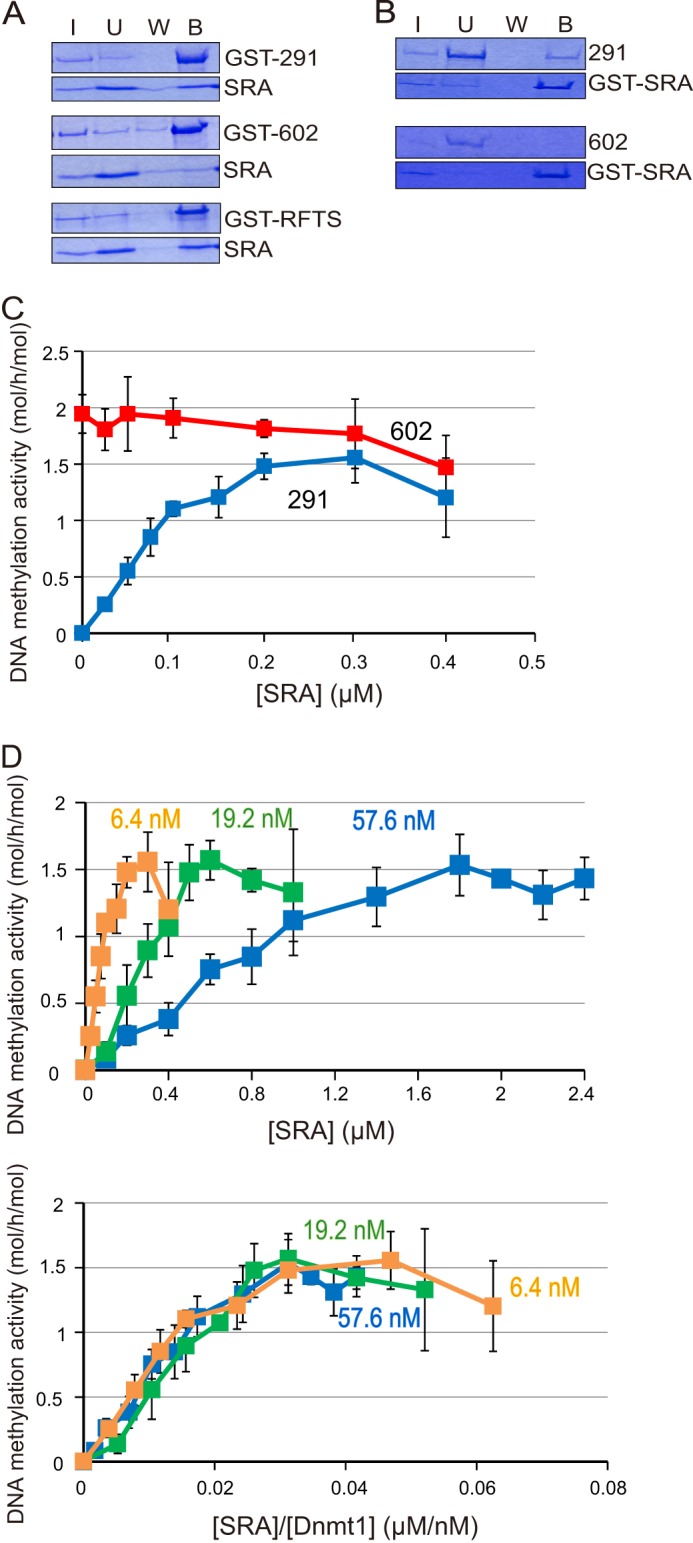
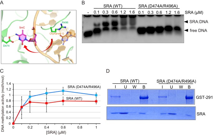
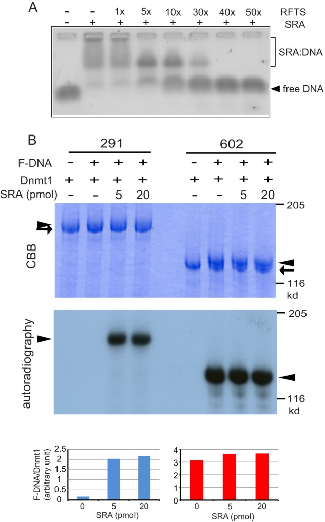
Dnmt1 is responsible for the maintenance DNA methylation during replication to propagate methylation patterns to the next generation. The replication foci targeting sequence (RFTS), which plugs the catalytic pocket, is necessary for recruitment of Dnmt1 to the replication site. In the present study we found that the DNA methylation activity of Dnmt1 was DNA length-dependent and scarcely methylated 12-bp short hemi-methylated DNA. Contrarily, the RFTS-deleted Dnmt1 and Dnmt1 mutants that destroyed the hydrogen bonds between the RFTS and catalytic domain showed significant DNA methylation activity even toward 12-bp hemi-methylated DNA. The DNA methylation activity of the RFTS-deleted Dnmt1 toward 12-bp hemi-methylated DNA was strongly inhibited on the addition of RFTS, but to a lesser extent by Dnmt1 harboring the mutations that impair the hydrogen bond formation. The SRA domain of Uhrf1, which is a prerequisite factor for maintenance methylation and selectively binds to hemi-methylated DNA, stimulated the DNA methylation activity of Dnmt1. The SRA to Dnmt1 concentration ratio was the determinant for the maximum stimulation. In addition, a mutant SRA, which had lost the DNA binding activity but was able to bind to Dnmt1, stimulated the DNA methylation activity of Dnmt1. The results indicate that the DNA methylation activity of Dnmt1 was stimulated on the direct interaction of the SRA and Dnmt1. The SRA facilitated acceptance of the 12-bp fluorocytosine-containing DNA by the catalytic center. We propose that the SRA removes the RFTS plug from the catalytic pocket to facilitate DNA acceptance by the catalytic center.

Keywords: DNA Methylation; DNA Methyltransferase; Epigenetics; Gene Regulation; Gene Silencing.


Figures

FIGURE 1.
FIGURE 1.
DNA length-dependent DNA methylation activities of Dnmt1(291–1620) and Dnmt1(602–1620). A, schematic illustrates Dnmt1. Dnmt1 has a multidomain structure (4, 14). The Dnmt1 constructs, Dnmt1(291–1620) (291) and Dnmt1(602–1620) (602) used in this study are indicated. B, DNA length-dependent DNA methylation activity of Dnmt1(291–1620) (left panel, 291) and Dnmt1(602–1620) (right panel, 602) toward hemi-methylated DNA of 12, 16, 20, 30, and 42 bp with one hemi-methylated CpG is shown. C, Dnmt1(602–1620) was incubated with the RFTS, and then the DNA methylation activity toward 12-bp (blue) and 42-bp hemi-methylated DNA (red) was determined. Specific activities of DNA methylation (mol of CH3 transferred/h/mol of Dnmt1) were determined. The average activities ± S.D. (error bars; n = 3) are shown. The inhibition of Dnmt1(602–1620) was statistically significant at 50, 100, and 200 nm RFTS (p < 0.001).
FIGURE 2.
FIGURE 2.
Dnmt1(291–1620), which disrupted the hydrogen bonds between the RFTS and catalytic domains, exhibits significant DNA methylation activity toward 12-bp hemi-methylated DNA. A, image of the four hydrogen bonds between the RFTS (light violet) and catalytic domains (light blue) of Dnmt1 (taken from Protein Data Bank ID code 3AV6). The hydrogen bonds between the side chains of Glu-531, Asp-532, and Asp-554, and the main chain of Leu-593 in the RFTS and Lys-1537, Arg-1576, Ser-1495, and Thr-1505, respectively, are shown. B, DNA methylation activities of Dnmt1(291–1620) (291), Dnmt1(602–1620) (602), and Dnmt1(291–1620) with E531A/D532A, and D554K determined at 37, 30, 25, and 20 °C (bars from left to right), respectively, toward 42-bp DNA with 12 hemi-methylated CpG. Average activities ± S.D. (error bars; n = 3) are shown (left panel). The logarithms of DNA methylation activities obtained (ln(DNA methylation activity), ordinate axis) against inverse temperatures (1/T, abscissa) are plotted (Arrhenius plot) (right panel). C, DNA methylation activities of Dnmt1(291–1620) (291), Dnmt1(602–1620) (602), and Dnmt1(291–1620) with the mutations E531A and D532A (E531A/D532A), D554A (D554K), and E531K and D532A (E531A/D532K) determined toward 12-bp hemi-methylated DNA as in Fig. 1. D, Dnmt1(602–1620) incubated with the RFTS domain without any mutation (WT, red) or with mutation E531A/D532A (dark green) or D544K (light green). Then the DNA methylation activity toward 12-bp hemi-methylated DNA was determined. The average values ± S.D. (n = 3) are plotted. E, the indicated amounts of the RFTS incubated with 12-bp or 42-bp DNA with one hemi-methylated CpG (0.2 μm). After the incubation, the mixtures were subjected to gel electrophoresis, and then DNA was visualized.
FIGURE 3.
FIGURE 3.
The SRA binds to the RFTS of Dnmt1 and induces DNA methylation activity toward 12-bp hemi-methylated DNA. A, the SRA was incubated with GST-tagged Dnmt1(291–1620) (GST-291), Dnmt1(602–1620) (GST-602), or Dnmt1(291–602) (GST-RFTS) bound to GSH-Sepharose. After the incubation, input (I), unbound (U), wash (W), and bound (B) fractions were analyzed by SDS-polyacrylamide gel electrophoresis. The protein bands were visualized by CBB staining. Equivalent amounts of samples were loaded, other than for the input (I) fractions, the amounts loaded being one fourth of those of the other fractions. B, Dnmt1(291–1620) (291) or Dnmt1(602–1620) (602) was incubated with GST-tagged SRA (GST-SRA) bound to GSH-Sepharose. Input (I), unbound (U), wash (W), and bound (B) fractions were analyzed as in A. C, DNA methylation activities of Dnmt1(291–1620) (blue) and Dnmt1(602–1620) (red) toward 12-bp hemi-methylated DNA were titrated with the SRA. The specific activities of DNA methylation (mol of CH3 transferred/h/mol of Dnmt1) were determined, and the average activities ± S.D. (error bars; n = 3) are shown. The stimulation of the DNA methylation activities of Dnmt1(291–1620) was statistically significant at all of the SRA concentrations examined (p < 0.001), except for 0.05 μm SRA where p < 0.01. D, DNA methylation activities toward 12-bp hemi-methylated DNA were determined by titrating the SRA with a fixed amount of DNA and three different concentrations of Dnmt1(291–1620): 6.4 (light brown), 19.2 (light green), and 57.6 nm (blue). Average activities ± S.D. (n = 3) are shown (upper panel). The concentrations of SRA in the upper panel were normalized with Dnmt1 and the DNA methylation activities are replotted (lower panel).
FIGURE 4.
FIGURE 4.
The mutant SRA as the amino acid residues responsible for flipping of the methylated cytosine out of double-stranded DNA cannot bind to 12-bp hemi-methylated DNA but can stimulate DNA methylation activity. A, image of the structure of the SRA bound to hemi-methylated DNA and flipping out of the methylated cytosine (5mC) of the double-stranded DNA is shown. Asp-474 is holding 5mC, and Arg-496 is stabilizing the orphaned Gly. The red arrow indicates the flipping of 5mC. The structure data were taken from PDB accession number 2ZKD. B, gel-shift assaying of the SRA and that with D474A/R496A was performed as in Fig. 2E. 12-bp hemi-methylated DNA (0.2 μm) was incubated with the indicated amounts of the SRA or the mutant. Free DNA (arrows) and DNA-bound SRA (arrowheads) are shown. C, DNA methylation activities of Dnmt1(291–1620) (12.8 μm) toward 12-bp hemi-methylated DNA were determined by titrated with SRA (WT, red) or SRA(D474A/R496A, blue). The average activities ± S.D. (n = 4) are shown. The stimulation of the DNA methylation activities of Dnmt1(291–1620) was statistically significant at all of the SRA (D474A/R496A) concentrations examined (p < 0.001), except for 0.1 μm SRA (D474A/R496A) where p < 0.05. Error bars, S.D. D, the SRA or mutant was incubated with GST-tagged Dnmt1(291–1620) (GST-291) bound to GSH-Sepharose. After the incubation, input (I), unbound (U), wash (W), and bound (B) fractions were analyzed by SDS-polyacrylamide gel electrophoresis. The protein bands were visualized by CBB staining. Equivalent amounts of samples were loaded, other than for the input (I) fractions, the amounts loaded being one fourth of those of the other fractions.
FIGURE 5.
FIGURE 5.
Interaction between the SRA and RFTS domains facilitates accession of 12-bp hemi-methylated DNA to the catalytic center of Dnmt1. A, the SRA binding to 12-bp hemi-methylated DNA was competed for by the RFTS in a dose-dependent manner. SRA (0.6 μm) and 12-bp hemi-methylated DNA (0.2 μm) binding was competed for with 1–50-fold higher concentrations of RFTS and then analyzed by gel shift assaying. B, Dnmt1(291–1620) or Dnmt1(602–1620) was incubated with 12-bp 32P-labeled F-DNA in the absence or presence of the SRA. After the reaction, samples were subjected to SDS-polyacrylamide gel electrophoresis, and the protein bands and radiolabeled F-DNA were visualized with CBB (top panel) and autoradiography (middle panel), respectively. Arrows and arrowheads indicate free Dnmt1 and Dnmt1 bound to F-DNA, respectively. The radioactive bands visualized with a BAS2000 were quantitated and normalized to those of the CBB-stained Dnmt1 bands (bottom panel).

References

    1. Li E. (2002) Chromatin modification and epigenetic reprogramming in mammalian development. Nat. Rev. Genet. 3, 662–673 - PubMed
    1. Reik W., Lewis A. (2005) Co-evolution of X chromosome inactivation and imprinting in mammals. Nat. Rev. Genet. 6, 403–410 - PubMed
    1. Goll M. G., Bestor T. H. (2005) Eukaryotic cytosine methyltransferases. Annu. Rev. Biochem. 74, 481–514 - PubMed
    1. Suetake I., Hayata D., Tajima S. (2006) The amino terminus of mouse DNA methyltransferase 1 forms an independent domain and binds to DNA with the sequence involving PCNA binding motif. J. Biochem. 140, 763–776 - PubMed
    1. Rountree M. R., Bachman K. E., Baylin S. B. (2000) DNMT1 binds HDAC2 and a new co-repressor, DMAP1, to form a complex at replication foci. Nat. Genet. 25, 269–277 - PubMed

Publication types

MeSH terms

LinkOut - more resources