

Structural features of glycol-split low-molecular-weight heparins and their heparin lyase generated fragments
- PMID: 24253408
- PMCID: PMC3925387
- DOI: 10.1007/s00216-013-7446-4
Structural features of glycol-split low-molecular-weight heparins and their heparin lyase generated fragments
Abstract
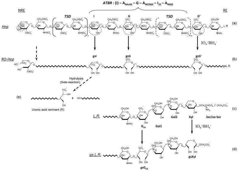
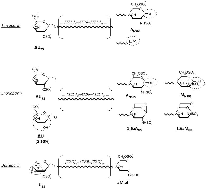
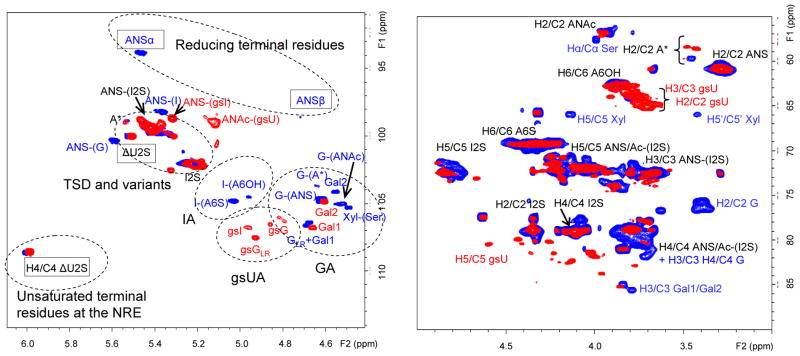
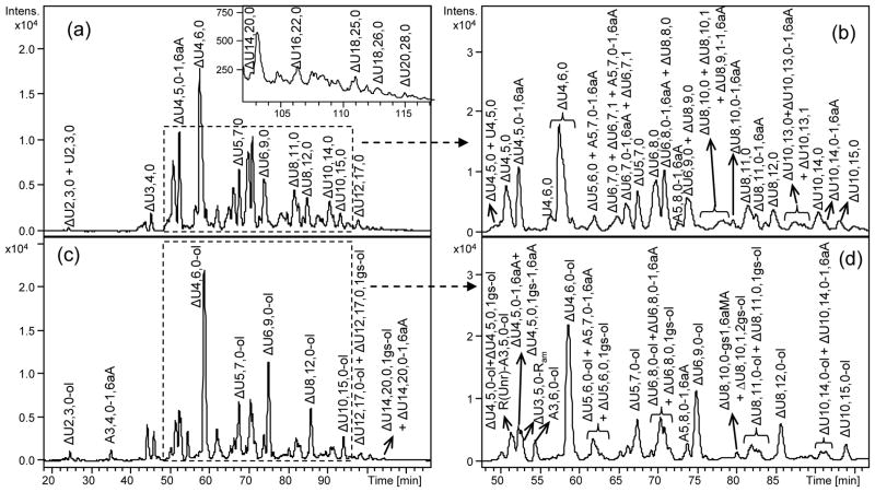
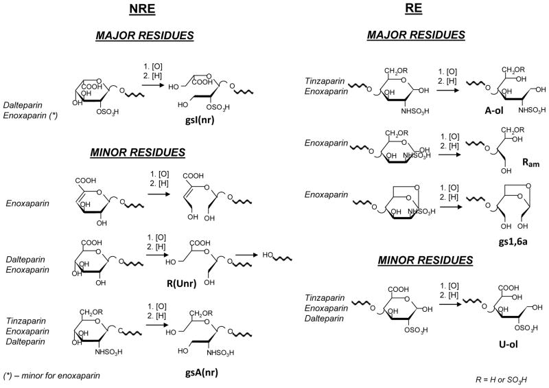
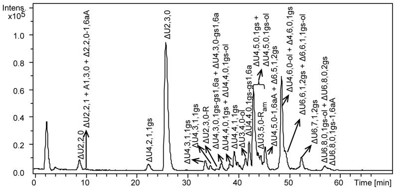
Periodate oxidation followed by borohydride reduction converts the well-known antithrombotics heparin and low-molecular-weight heparins (LMWHs) into their "glycol-split" (gs) derivatives of the "reduced oxyheparin" (RO) type, some of which are currently being developed as potential anti-cancer and anti-inflammatory drugs. Whereas the structure of gs-heparins has been recently studied, details of the more complex and more bioavailable gs-LMWHs have not been yet reported. We obtained RO derivatives of the three most common LMWHs (tinzaparin, enoxaparin, and dalteparin) and studied their structures by two-dimensional nuclear magnetic resonance spectroscopy and ion-pair reversed-phase high-performance liquid chromatography coupled with electrospray ionization mass spectrometry. The liquid chromatography-mass spectrometry (LC-MS) analysis was extended to their heparinase-generated oligosaccharides. The combined NMR/LC-MS analysis of RO-LMWHs provided evidence for glycol-splitting-induced transformations mainly involving internal nonsulfated glucuronic and iduronic acid residues (including partial hydrolysis with formation of "remnants") and for the hydrolysis of the gs uronic acid residues when formed at the non-reducing ends (mainly, in RO-dalteparin). Evidence for minor modifications, such as ring contraction of some dalteparin internal aminosugar residues, was also obtained. Unexpectedly, the N-sulfated 1,6-anhydromannosamine residues at the enoxaparin reducing end were found to be susceptible to the periodate oxidation. In addition, in tinzaparin and enoxaparin, the borohydride reduction converts the hemiacetalic aminosugars at the reducing end to alditols. Typical LC-MS signatures of RO-derivatives of individual LMWH both before and after digestion with heparinases included oligosaccharides generated from the original antithrombin-binding and "linkage" regions.
Figures
Similar articles
-
Susceptibility of enoxaparin reducing end amino sugars to periodate oxidation.Carbohydr Res. 2014 Dec 5;400:33-43. doi: 10.1016/j.carres.2014.08.016. Epub 2014 Sep 10. Carbohydr Res. 2014. PMID: 25457608 Free PMC article.
-
Profiling glycol-split heparins by high-performance liquid chromatography/mass spectrometry analysis of their heparinase-generated oligosaccharides.Anal Biochem. 2013 Mar 1;434(1):112-22. doi: 10.1016/j.ab.2012.11.011. Epub 2012 Nov 29. Anal Biochem. 2013. PMID: 23201389 Free PMC article.
-
Characterization of Low-Molecular-Weight Heparins by Strong Anion-Exchange Chromatography.J AOAC Int. 2017 Nov 1;100(6):1706-1714. doi: 10.5740/jaoacint.17-0217. Epub 2017 Jul 13. J AOAC Int. 2017. PMID: 28707621
-
Differentiation of the low-molecular-weight heparins.Pharmacotherapy. 2001 Jun;21(6 Pt 2):62S-70S; discussion 71S-72S. doi: 10.1592/phco.21.8.62s.34594. Pharmacotherapy. 2001. PMID: 11401195 Review.
-
Structural modification induced in heparin by a Fenton-type depolymerization process.Semin Thromb Hemost. 2007 Jul;33(5):466-77. doi: 10.1055/s-2007-982077. Semin Thromb Hemost. 2007. PMID: 17629843 Review.
Cited by
-
Structural Characterization of the Low-Molecular-Weight Heparin Dalteparin by Combining Different Analytical Strategies.Molecules. 2017 Jun 24;22(7):1051. doi: 10.3390/molecules22071051. Molecules. 2017. PMID: 28672818 Free PMC article.
-
The Low Molecular Weight Heparin Tinzaparin Attenuates Platelet Activation in Terms of Metastatic Niche Formation by Coagulation-Dependent and Independent Pathways.Molecules. 2018 Oct 24;23(11):2753. doi: 10.3390/molecules23112753. Molecules. 2018. PMID: 30356007 Free PMC article.
-
Targeting heparin and heparan sulfate protein interactions.Org Biomol Chem. 2017 Jul 21;15(27):5656-5668. doi: 10.1039/c7ob01058c. Epub 2017 Jun 27. Org Biomol Chem. 2017. PMID: 28653068 Free PMC article. Review.
-
Binding ability of methylene blue with heparin dependent on its sulfate level rather than its sulfation location or basic saccharide structure.Glycoconj J. 2021 Oct;38(5):551-560. doi: 10.1007/s10719-021-10010-2. Epub 2021 Sep 13. Glycoconj J. 2021. PMID: 34515908
-
Heparanase as an Additional Tool for Detecting Structural Peculiarities of Heparin Oligosaccharides.Molecules. 2019 Dec 2;24(23):4403. doi: 10.3390/molecules24234403. Molecules. 2019. PMID: 31810297 Free PMC article.
References
-
- Weitz JI. Low-molecular weight heparins. N Engl J Med. 1997;337:688–698. - PubMed
-
- Guerrini M, Guglieri S, Naggi A, Sasisekharan R, Torri G. Low molecular weight heparins: structural differentiation by bidimentional nuclear magnetic resonance spectroscopy. Semin Thromb Hemost. 2007;33:478–487. and references therein. - PubMed
-
- Casu B. In: Chemistry and Biology of Heparin and Heparan Sulfate. Garg HG, Linhardt RJ, Hales CA, editors. Elsevier; Amsterdam: 2005. and references therein.
-
- Lever R, Page CP. Novel drug development opportunities for heparin. Nat Rev Drug Discov. 2002;1:140–148. - PubMed
-
- Fuster MM, Esko JD. The sweet and sour of cancer: glycans as novel therapeutic agents. Nat Rev Cancer. 2005;5:526–542. - PubMed
Publication types
MeSH terms
Substances
Grants and funding
LinkOut - more resources
Full Text Sources
Other Literature Sources
Research Materials
