. 2013:4:2770.
doi: 10.1038/ncomms3770.

Continent-wide panmixia of an African fruit bat facilitates transmission of potentially zoonotic viruses

Affiliations

Continent-wide panmixia of an African fruit bat facilitates transmission of potentially zoonotic viruses

Alison J Peel et al. Nat Commun. 2013.

Abstract

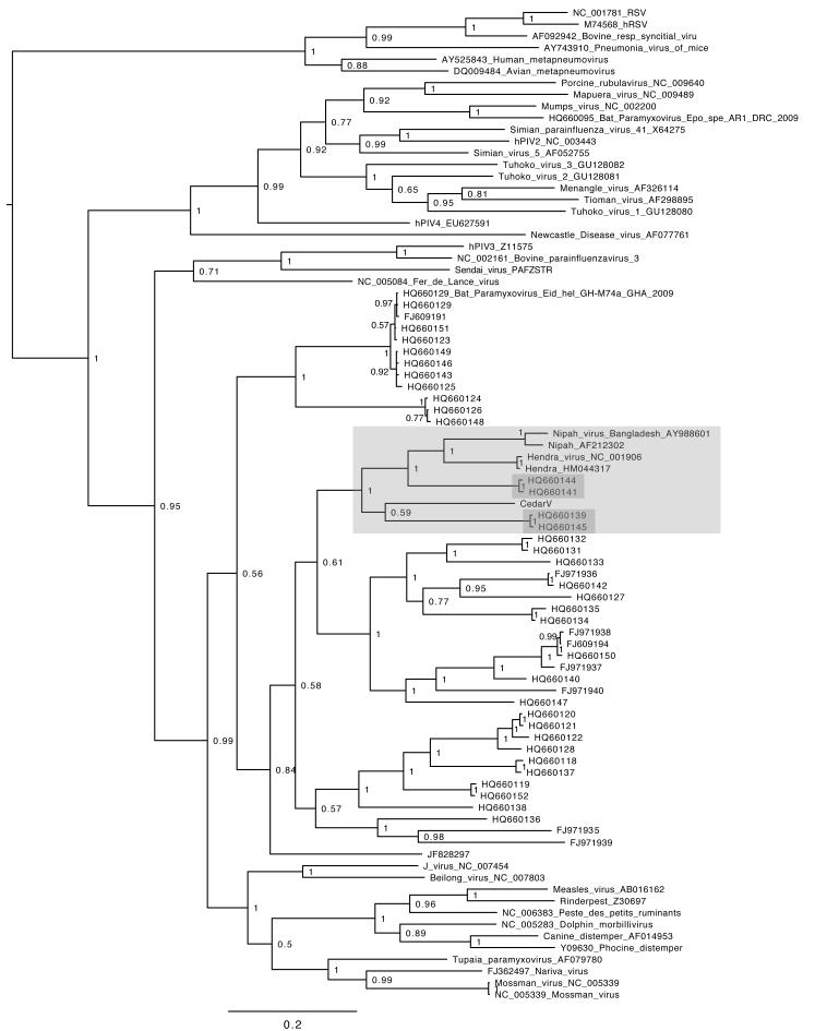
The straw-coloured fruit bat, Eidolon helvum, is Africa's most widely distributed and commonly hunted fruit bat, often living in close proximity to human populations. This species has been identified as a reservoir of potentially zoonotic viruses, but uncertainties remain regarding viral transmission dynamics and mechanisms of persistence. Here we combine genetic and serological analyses of populations across Africa, to determine the extent of epidemiological connectivity among E. helvum populations. Multiple markers reveal panmixia across the continental range, at a greater geographical scale than previously recorded for any other mammal, whereas populations on remote islands were genetically distinct. Multiple serological assays reveal antibodies to henipaviruses and Lagos bat virus in all locations, including small isolated island populations, indicating that factors other than population size and connectivity may be responsible for viral persistence. Our findings have potentially important public health implications, and highlight a need to avoid disturbances that may precipitate viral spillover.


Figures

Fig 1
Fig 1. Map showing location of E. helvum sampling locations for genetic and serological analyses
Shading represents the distribution range of E. helvum. Sampling locations are numbered as in Supplementary Data 1. Adapted from Mickleburgh et al. .
Fig 2
Fig 2. Isolation by distance plots of pairwise population values for log geographic distance and genetic distance
Genetic distance is given by Slatkin’s linearised φST (φST /(1- φST) for cytochrome b mtDNA analyses (left column) or Slatkin’s linearised FST (FST/(1-FST) for microsatellite analyses (right column). Note that the scales vary. Analyses were performed for all E. helvum populations (n = 12), for continental populations only (n = 9), or for island populations only (n=4). Statistical significance was assessed using a Mantel test and p-values are shown where sample size was sufficient to allow testing. Geographic distance is given in km.
Fig 3
Fig 3. Eidolon helvum cytochrome b median joining haplotype network
No spatial clustering is present in continental African countries or within regions. Each circle represents a unique haplotype, and its size is proportional to its frequency. Lines represent base pair changes between two haplotypes, with the length proportional to the number of base pair changes. Main haplotypes and those containing island samples are labelled by name. Inset in the bottom right shows the relationship between the haplotype network and three clades identified in the Bayesian phylogeny.
Fig 4
Fig 4. Estimated population structure
Estimates from STRUCTURE analyses for K = 2 to 5 based on microsatellite data from 502 individuals. Analyses run using the admixture setting identified three clusters corresponding to continental and Bioko populations (left), São Tomé and Príncipe (centre, orange) and Annobón (right, red). Each vertical line represents the proportional membership assignment of one individual to each of K coloured clusters. Black lines divide the plot into sampling locations. Ghana (GH), DRC (DR), Kenya (KE), Zambia (ZA), Malawi (MA), Tanzania (TZ), Uganda (UG), Rio Muni (RM), Bioko (BI), Príncipe (PR), São Tomé (ST), Annobón (AN).
Fig 5
Fig 5. Diversity of paramyxoviruses in Eidolon helvum urine collected across multiple African sites detected using Paramyxovirinae-targeted PCR
Phylogenetic tree for a 531 bp segment of the polymerase gene of members of the subfamily Paramyxovirinae, including sequences generated in this study and publicly available paramyxovirus sequences (with GenBank accession numbers). Relevant posterior probability values are shown. Horizontal branches are drawn to a scale of nucleotide substitutions per site. Individual extraction pools IDs are followed by letters denoting the clone. Groups containing sequences previously uncharacterized sequences that display a common phylogenetic origin supported by high posterior probability values (≥0.95) are highlighted by numbered light grey boxes. Within these boxes, sequences obtained from samples collected from Tanzania and Uganda are further highlighted by darker grey boxes. Pair wise nucleotide identities of the sequences from samples collected Tanzania and Uganda with their nearest phylogenetic relative are shown within the grey boxes. One PCR-positive Ugandan pooled sample (sample 23) contained paramyxoviral sequence with 95% nucleotide sequence identity with sequences detected in Ghana that comprised part of a phylogenetically-distinct lineage of unclassified bat-derived viruses (group 5). Of the two PCR-positive Tanzanian samples, one contained paramyxoviral sequence related to mumps virus (sample 21) and shared 98% nucleotide identity with a Ghanaian sequence (group 2), and the other (sample 13) contained a sequence related to, but distinct from (74% nucleotide identity) sequences detected in Ghana (group 3).
Fig 6
Fig 6. Henipavirus phylogenetic relationships
Phylogeny based on a 559 bp segment of the polymerase gene incorporating fragments known Paramyxovirinae and fragments from Drexler et al. The clade containing known henipaviruses (Hendra virus (HeV), Nipah Virus (NiV) and Cedar virus (CedPV)) is highlighted in pale gray. Sequence fragments from viruses detected in E. helvum within this clade are further highlighted by dark gray boxes. Posterior probability values are shown and the bar represents 0.2 expected nucleotide substitutions per site. GenBank accession numbers are shown.

Comment in

Similar articles

Cited by

References

    1. Hayman DT, et al. Ecology of Zoonotic Infectious Diseases in Bats: Current Knowledge and Future Directions. Zoonoses Public Hlth. 2013;60(1):2–21. doi: 10.1111/zph.12000. - PMC - PubMed
    1. Banyard AC, Hayman DTS, Johnson N, McElhinney LM, Fooks AR. Bats and lyssaviruses. Adv. Virus Res. 2011;79:239–289. - PubMed
    1. Hayman DTS, et al. Evidence of Henipavirus Infection in West African Fruit Bats. PLoS ONE. 2008;3:e2739. - PMC - PubMed
    1. Mickleburgh S, Hutson A, Bergmans W, Fahr J, Racey PA. Eidolon helvum. In: IUCN, editor. 2008 IUCN Red List of Threatened Species. 2008. Available from: www.iucnredlist.org. Downloaded on 6 February 2011.
    1. Hayman DTS, et al. Endemic Lagos bat virus infection in Eidolon helvum. Epidemiol Infect. 2012;140:2163–2171. - PMC - PubMed

Publication types

Substances