. 2013:4:2773.
doi: 10.1038/ncomms3773.

Platelet-targeted gene therapy with human factor VIII establishes haemostasis in dogs with haemophilia A

Affiliations
Free PMC article

Platelet-targeted gene therapy with human factor VIII establishes haemostasis in dogs with haemophilia A

Lily M Du et al. Nat Commun. 2013.
Free PMC article

Abstract

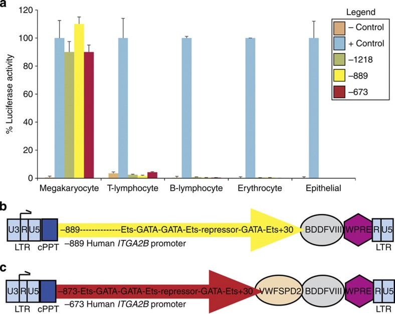
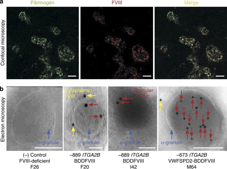
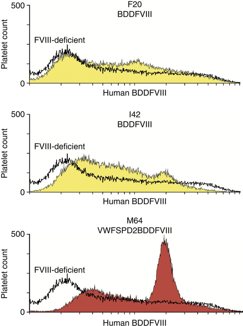
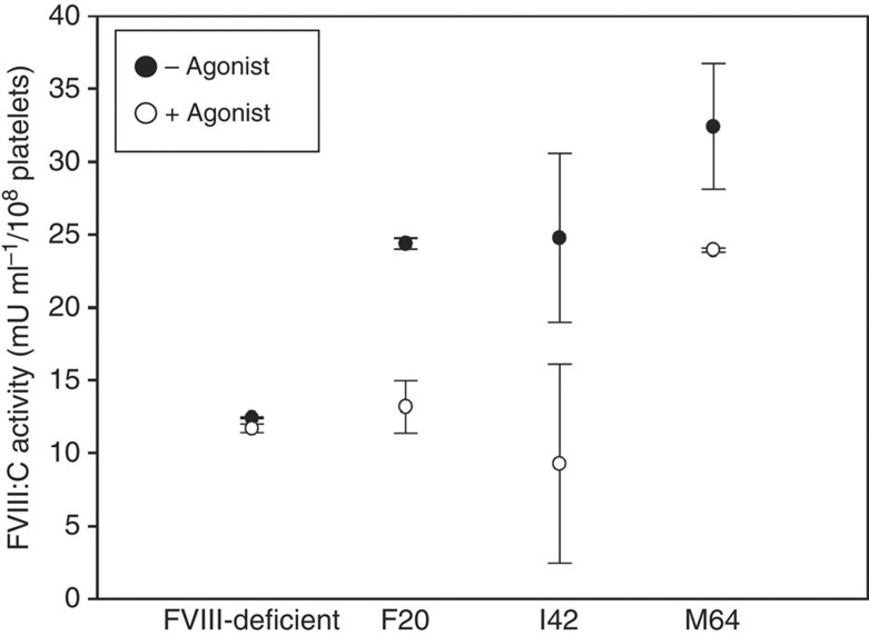
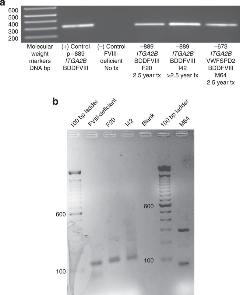
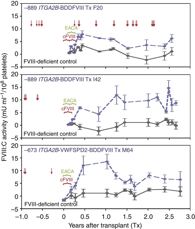
It is essential to improve therapies for controlling excessive bleeding in patients with haemorrhagic disorders. As activated blood platelets mediate the primary response to vascular injury, we hypothesize that storage of coagulation Factor VIII within platelets may provide a locally inducible treatment to maintain haemostasis for haemophilia A. Here we show that haematopoietic stem cell gene therapy can prevent the occurrence of severe bleeding episodes in dogs with haemophilia A for at least 2.5 years after transplantation. We employ a clinically relevant strategy based on a lentiviral vector encoding the ITGA2B gene promoter, which drives platelet-specific expression of human FVIII permitting storage and release of FVIII from activated platelets. One animal receives a hybrid molecule of FVIII fused to the von Willebrand Factor propeptide-D2 domain that traffics FVIII more effectively into α-granules. The absence of inhibitory antibodies to platelet-derived FVIII indicates that this approach may have benefit in patients who reject FVIII replacement therapies. Thus, platelet FVIII may provide effective long-term control of bleeding in patients with haemophilia A.


Conflict of interest statement

David A. Wilcox and Sandra L. Haberichter have applied for a US Provisional Patent Application (serial no. 61/717,951) entitled “Platelet Targeted Treatment” for the therapy described within this manuscript. All other authors declare no competing interests.

Figures

Figure 1
Figure 1. Platelet-targeted lentiviral vector design.
(a) ITGA2B gene promoter fragments direct megakaryocyte-specific expression in luciferase reporter assay. Human pro-megakaryocytic, lymphocytic, erythrocytic and epithelial cell lines are transfected with a luciferase reporter construct under the control of one of three fragments of the human ITGA2B gene promoter beginning at either nucleotide −1218(green), −889(yellow) or −673(red) in-frame with the luciferase gene. A luciferase construct without a gene promoter served as a negative (−)control (peach), whereas a construct with a tissue-nonspecific gene promoter of the cytomegalovirus (CMV) is a positive (+)control (blue) arbitrarily assigned a value of 100% luciferase activity. Lysates of each cell line (x axis) were measured in duplicate on a luminometer to report the percent (%) luciferase activity (y axis) for each construct within each cell line. Cells were co-transfected with plasmid constructs encoding the marker gene, β-galactosidase, under the under the control of the CMV promoter to correct for variations in transfection efficiency. Shown is one representative of nine experiments where the error bars represent the s.d. from the mean value of measurements made in duplicate. (b) −889ITGA2B-BDDFVIII-WPTS lentiviral vector diagram. Shown is the region between the 5′-long terminal repeat (LTR) (left, blue box) and 3′-LTR without a U3 enhancer/promoter (right, blue box) to permit the ITGA2B promoter (yellow arrow) to direct megakaryocyte-specific synthesis of BDDFVIII (grey circle). The −889ITGA2B promoter binds GATA and Ets transcription factors for high-level gene expression in megakaryocytes and a repressor region inhibits gene expression in other lineages (yellow arrow). The central polypurine tract (cPPT, navy) and woodchuck hepatitis virus postregulatory element (WPRE, purple diamond) enhance efficiency of transgene expression. (c) −673ITGA2B-VWFSPD2-BDDFVIII-WPTS lentiviral vector diagram. The backbone of this vector is the same as described in b, except a smaller −673ITGA2B promoter (red arrow) was fused to DNA encoding the human VWF signal peptide (SP) and D2 domain (peach circle) to traffic BDDFVIII (grey circle) directly to platelet α–granules.
Figure 2
Figure 2. Synthesis and trafficking of BDDFVIII into canine platelet α-granules.
(a) Confocal microscopy showing co-localization of BDDFVIII and Fg within platelets. Canine CD34+G-PBC were transduced with lentivirions encoding human BDDFVIII followed by transplant haemophilia A dogs. Peripheral blood platelets were isolated from whole blood, fixed, permeabilized and examined by indirect immunofluorescence analysis for Fg and BDDFVIII distribution. Shown is a representative image using a × 10 eye piece and × 100 oil objective and a × 2 digital zoom of platelets isolated from one transplanted animal (I42) from an experiment that was performed seven times on all three dogs and a FVIII-deficient negative control. Fg was visualized with a 1°Ab to this platelet-specific marker for α-granules and a Alexa488-conjugated 2°Ab (left, green). BDDFVIII was detected with 1°Ab to human FVIII and an Alexa568-conjugated 2°Ab (middle, red). BDDFVIII colocalized with Fg is observed when the two images are merged (right, yellow). The white scale bar is 5 μm in length. (b) Electron microscopy localized human BDDFVIII directly in α-granules. Shown is a representative image of BDDFVIII absent from an ultrathin cryosection of a single platelet α-granule (blue arrow) from a FVIII-deficient (negative control, F26) when probed with a 1°Ab (301.3) to human BDDFVIII and a 2°Ab-conjugated to 10-nm gold particles. In contrast, BDDFVIII was localized directly within the α-granule (red arrow) and within intracytoplasmic membrane systems (yellow arrow) in representative images from all three experimental dogs (F20, I42 and M64). Interestingly, sections from dog (M64) receiving the −673ITGA2B-VWFSPD2-BDDFVIII-transduced G-PBC (VWF-targeting peptide) exhibited a noticably higher concentration of BDDFVIII within mature platelet α-granules. Each sample was cut on a minimum of three separate occasions with a series of ultrathin sections subjected to immunogold labelling and analysis of at least 100 α-granules per sample. The white scale bar is 0.2 μm in length.
Figure 3
Figure 3. Quantitative analysis of platelet FVIII.
Flow cytometric analysis of platelets. BDDFVIII was absent in platelets (x axis) analysed from a FVIII-deficient dog used as a negative control for staining with a human BDDFVIII 1°Ab and Alexa Fluor 568-conjugated 2°Ab (black, unshaded histogram). In contrast G-PBC from transplanted dogs displayed appreciable levels of platelet BDDFVIII (shaded histogram: −889ITGA2B-driven FVIII of F20 and I42, yellow; −673ITGA2B-driven FVIII of M64, red). The hierarchy for the mean fluorescence intensity of FVIII expression reveals that F20<I42<M64. Shown are the results from one experiment analysis of 50,000 platelets per sample at 2.9 (F20), 1.9 (I42) and 0.9 (M64) years after infusion of FVIII-transduced G-PBC, which is representative of the outcome of seven separate experiments.
Figure 4
Figure 4. Activated platelets induced to secrete FVIII:C.
To detect FVIII:C activity present in lysate of quiescent, untreated platelets (black circle; (−) agonist) and FVIII:C activity remaining in lysate after secretion of BDDFVIII from platelets treated with a mixture of physiological agonists of platelet activation: ADP, epinephrine and canine PAR1,3,4 (white circle: (+) agonist). Platelet lysates from a FVIII-deficient negative control dog show that the level of FVIII:C background activity is virtually unchanged for untreated and activated platelets. Dogs that received BDDFVIII-transduced G-PBC show an appreciable decrease in FVIII:C activity only after platelet activation; thus demonstrating that platelets isolated from experimental dogs can be induced to secrete FVIII. Each data point represents the mean value of FVIII:C activity from two independent samples each measured in duplicate with s.d. represented by black error bars. Shown is the result from one experiment performed at 2.7 (F20), 1.6 (I42) and 0.6 (M64) years after infusion of FVIII-transduced G-PBC, which is representative of the outcome of three separate experiments.
Figure 5
Figure 5. PCR analysis for detection and localization of lentiviral vector within canine genome.
(a) Long-term detection of BDDFVIII-lentiviral vector within leukocyte genomic DNA. Shown is an ethidium bromide-stained agarose gel of PCR product-derived genomic DNA isolated from canine peripheral blood leukocytes. Molecular weight markers (Lane 1) are labelled on the left in base pairs (bp). In Lane 2, p−889ITGA2B-BDDFVIII-WPTS served as a positive control for PCR of a 302 bp region of the lentiviral WPRE. PCR failed to detect WPRE within leukocyte genomic DNA from a FVIII-deficient dog serving as a negative control (Lane 3). In contrast, WPRE was detected in the PCR of all three experimental dogs (F20, I42, M64; Lanes 4–6) for at least 2.5 years after G-PBC transplant demonstrating long-term insertion of the lentiviral vector into the canine genome. All reactions were run in triplicate from at least three separate samples collected periodically for at least 2.5 years after G-PBC transplant. (b) Linear amplification-mediated (LAM)-PCR to localize lentiviral vector within canine genome. Polyacrylamide gel of LAM-PCR products from a FVIII-deficient negative control and experimental dogs at 2.5 (F20), 1.4 (I42) and 0.5 (M64) years after infusion of FVIII-transduced G-PBC. PCR was performed on genomic DNA isolated from leukocytes purified with Ficoll from circulating peripheral blood. Each amplified band represents a distinct proviral integration. All lanes appear to contain multiple insertions, indicative of polyclonality. The panel underwent black–white inversion from the original digital image, and levels were globally adjusted to improve visibility of all bands. DNA standard markers (Lane 1 and 6) labelled on left in base pairs.
Figure 6
Figure 6. Correction of the canine haemophilia A phenotype with platelet BDDFVIII.
A Coatest SP4 FVIII chromagenic analysis assay was performed on lysates of 1 × 10⁸ washed peripheral blood platelets resuspended in 1 ml of lysis buffer from samples collected periodically for at least 2.5 years after transplant (x axis). The mean value of FVIII activity (mU ml per 108 platelets) is represented by the plotting of data points recorded from two independent samples (each measured in duplicate) with error bars showing ±s.d. for at least 12 time points (y axis). Analysis of platelet FVIII:C activity from a FVIII-deficient dog served as a negative control (black, solid line). Interestingly, the level of FVIII:C activity was detected above background levels at regular intervals (blue dashed line) for ≈2.5 years after transplant of BDDFVIII-transduced G-PBC (F20, top; I42, middle; M64, bottom panel). Note, each dog was administered daily injections of FVIII in the form of canine blood transfusions or cFVIII (red bracket) and EACA (green bracket) for uncontrolled bleeding for a short time after transplant (until haemocult tests were negative). As anticipated, each dogs required cFVIII supplements (red arrows) to help resolve severe bleeding episodes incurred before G-PBC transplant. Remarkably, only F20 (which had the lowest steady state level of FVIII:C activity) required cFVIII supplements after G-PBC transplant demonstrating platelet FVIII:C activity reached therapeutic levels in I42 and M64.

Similar articles

Cited by

References

    1. Leslie M. Cell biology. Beyond clotting: the powers of platelets. Science 328, 562–564 (2010). - PubMed
    1. Stonebraker J. S., Bolton-Maggs P. H., Soucie J. M., Walker I. & Brooker M. A study of variations in the reported haemophilia A prevalence around the world. Haemophilia 16, 20–32 (2010). - PubMed
    1. Berntorp E. & Shapiro A. D. Modern haemophilia care. Lancet 379, 1447–1456 (2012). - PubMed
    1. Lozier J. N. et al. The Chapel Hill hemophilia A dog colony exhibits a factor VIII gene inversion. Proc. Natl Acad. Sci. USA 99, 12991–12996 (2002). - PMC - PubMed
    1. Viel K. R. et al. Inhibitors of factor VIII in black patients with hemophilia. N. Engl. J. Med. 360, 1618–1627 (2009). - PMC - PubMed

Publication types