Review
. 2014 Jan-Feb;5(1):15-27.
doi: 10.4161/gmic.26854. Epub 2013 Oct 31.

Clostridium difficile binary toxin CDT: mechanism, epidemiology, and potential clinical importance

Affiliations
Review

Clostridium difficile binary toxin CDT: mechanism, epidemiology, and potential clinical importance

Dale N Gerding et al. Gut Microbes. 2014 Jan-Feb.

Abstract

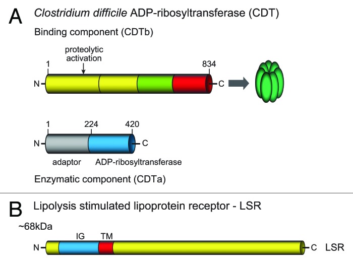
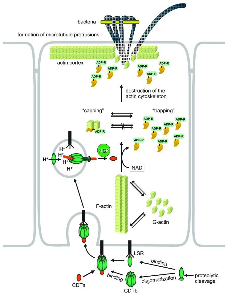
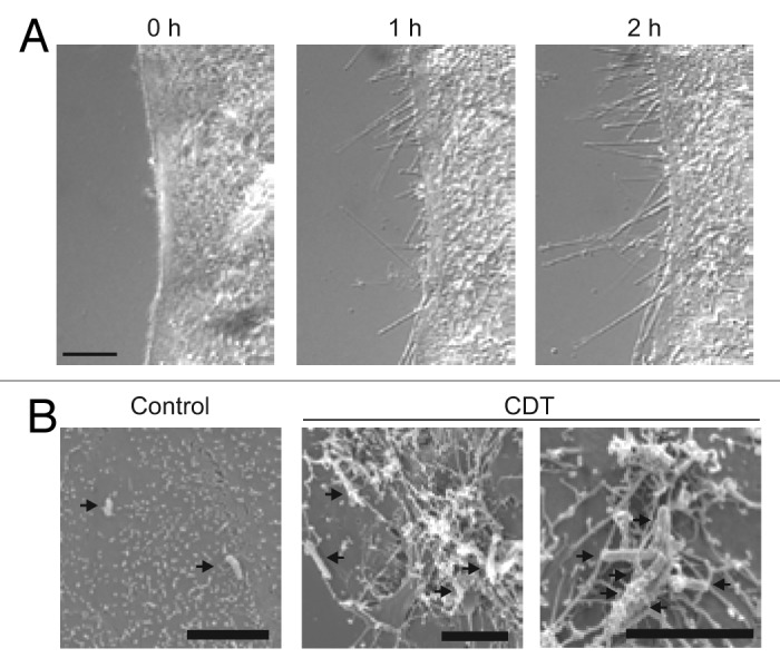
Binary toxin (CDT) is frequently observed in Clostridium difficile strains associated with increased severity of C. difficile infection (CDI). CDT belongs to the family of binary ADP-ribosylating toxins consisting of two separate toxin components: CDTa, the enzymatic ADP-ribosyltransferase which modifies actin, and CDTb which binds to host cells and translocates CDTa into the cytosol. CDTb is activated by serine proteases and binds to lipolysis stimulated lipoprotein receptor. ADP-ribosylation induces depolymerization of the actin cytoskeleton. Toxin-induced actin depolymerization also produces microtubule-based membrane protrusions which form a network on epithelial cells and increase bacterial adherence. Multiple clinical studies indicate an association between binary toxin genes in C. difficile and increased 30-d CDI mortality independent of PCR ribotype. Further studies including measures of binary toxin in stool, analyses of CDI mortality caused by CDT-producing strains, and examination of the relationship of CDT expression to TcdA and TcdB toxin variants and PCR ribotypes are needed.

Keywords: CDT; Clostridium difficileinfection; PCR ribotyping; binary toxin; disease severity; mechanism; mortality; toxinotyping.


Figures

None
Figure 1. Schematic representation of the CDT region and flanking genes. The regions from the nontoxigenic isolate CD37 (A), the binary toxin-negative isolate strain 630 (B), and the binary toxin-positive isolate QCD-32 g58 (C) are shown. The positions of the 5′ flanking genes CD2601 and CD2602, the 3′ flanking gene trpS, the response regulator gene cdtR, and the CDT binary toxin-encoding genes cdtAB, or their pseudogenes, are shown. For each variant of the CDT region the positions of the 5′ and 3′ conserved boundaries are shown, and the size of the entire CdtLoc is indicated. The unique 68-bp sequence that is present in CD37 and other nontoxigenic isolates in place of the CdtLoc is shown in bold. Adapted from J. Bacteriol. 2007;89:7290–7301, with permission from American Society for Microbiology.
None
Figure 2. Scheme of the structure of CDT. (A) The binary toxin consists of a binding component (CDTb) and an enzymatic component (CDTa). Both are expressed with leader sequences, which are not shown here. CDTb is activated by proteolytic cleavage to release ~20 and 75 kDa fragments. The 75 kDa fragment is the active binding component, which oligomerizes to form heptamers. (B) The lipolysis stimulated lipoprotein receptor (LRS) is the target cell receptor of CDT. The protein possesses an extracellular part with an immunoglobulin-like structure, a transmembrane region and a large intracellular part.
None
Figure 3. Up-take and mode of action of CDT. The proteolytically activated binding component of CDTb forms heptamers and binds to its cell surface receptor LSR. Alternatively, monomeric CDT binds to the receptor and, thereafter, polymerizes to form heptamers. Then, the enzymatic component CDTa interacts with CDTb. The LSR-CDT complex is endocytosed. At low pH of endosomes, the binding component inserts into the endosomal membrane and forms a pore. Through the pore, the enzymatic component is translocated into the cytosol. This process depends on cytosolic chaperon system, including heat shock proteins (HSP, cyclophillin A and FK506-binding protein 51). In the cytosol, CDTa ADP-ribosylates actin. ADP-ribosylated actin is not able to polymerize and is trapped in its monomeric form. Moreover, ADP-ribosylated actin acts like a capping protein to block polymerization at the barbed (plus) ends of F-actin. This causes enhanced depolymerization of the actin cytoskeleton. The depolymerization of cortical actin, which is located beneath the cell membrane, results in formation of long protrusions, which are microtubule based. The protrusions form a network on the surface of epithelial cells, which increases the interaction interface, and enhances adherence and colonization of clostridia.
None
Figure 4. Microscopic pictures of CDT-induced protrusions. (A) The microscopic pictures show protrusion formation after treatment of human carcinoma Caco-2 cells after CDT treatment. (B) Electron microscopic studies show clostridia embedded in the meshwork of protrusions (pictures are modified from Schwan et al.75).

References

    1. McDonald LC, Killgore GE, Thompson A, Owens RC, Jr., Kazakova SV, Sambol SP, Johnson S, Gerding DN. An epidemic, toxin gene-variant strain of Clostridium difficile. N Engl J Med. 2005;353:2433–41. doi: 10.1056/NEJMoa051590. - DOI - PubMed
    1. Loo VG, Poirier L, Miller MA, Oughton M, Libman MD, Michaud S, Bourgault AM, Nguyen T, Frenette C, Kelly M, et al. A predominantly clonal multi-institutional outbreak of Clostridium difficile-associated diarrhea with high morbidity and mortality. N Engl J Med. 2005;353:2442–9. doi: 10.1056/NEJMoa051639. - DOI - PubMed
    1. Goorhuis A, Bakker D, Corver J, Debast SB, Harmanus C, Notermans DW, Bergwerff AA, Dekker FW, Kuijper EJ. Emergence of Clostridium difficile infection due to a new hypervirulent strain, polymerase chain reaction ribotype 078. Clin Infect Dis. 2008;47:1162–70. doi: 10.1086/592257. - DOI - PubMed
    1. Rupnik M. Heterogeneity of large clostridial toxins: importance of Clostridium difficile toxinotypes. FEMS Microbiol Rev. 2008;32:541–55. doi: 10.1111/j.1574-6976.2008.00110.x. - DOI - PubMed
    1. Debast SB, van Leengoed LA, Goorhuis A, Harmanus C, Kuijper EJ, Bergwerff AA. Clostridium difficile PCR ribotype 078 toxinotype V found in diarrhoeal pigs identical to isolates from affected humans. Environ Microbiol. 2009;11:505–11. doi: 10.1111/j.1462-2920.2008.01790.x. - DOI - PubMed

Publication types

MeSH terms

Substances