. 2014 Mar;19(3):467-79.
doi: 10.1007/s10495-013-0944-1.

The mechanism by which MEK/ERK regulates JNK and p38 activity in polyamine depleted IEC-6 cells during apoptosis

Affiliations

The mechanism by which MEK/ERK regulates JNK and p38 activity in polyamine depleted IEC-6 cells during apoptosis

Mitul N Bavaria et al. Apoptosis. 2014 Mar.

Abstract

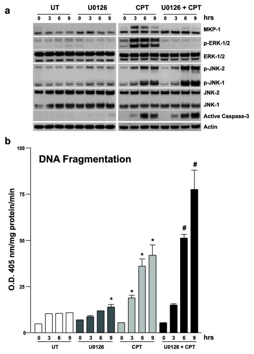
Polyamine-depletion inhibited apoptosis by activating ERK1/2, while, preventing JNK1/2 activation. MKP-1 knockdown by SiRNA increased ERK1/2, JNK1/2, and p38 phosphorylation and apoptosis. Therefore, we predicted that polyamines might regulate MKP1 via MEK/ERK and thereby apoptosis. We examined the role of MEK/ERK in the regulation of MKP1 and JNK, and p38 activities and apoptosis. Inhibition of MKP-1 activity with a pharmacological inhibitor, sanguinarine (SA), increased JNK1/2, p38, and ERK1/2 activities without causing apoptosis. However, pre-activation of these kinases by SA significantly increased camptothecin (CPT)-induced apoptosis suggesting different roles for MAPKs during survival and apoptosis. Inhibition of MEK1 activity prevented the expression of MKP-1 protein and augmented CPT-induced apoptosis, which correlated with increased activities of JNK1/2, caspases, and DNA fragmentation. Polyamine depleted cells had higher levels of MKP-1 protein and decreased JNK1/2 activity and apoptosis. Inhibition of MEK1 prevented MKP-1 expression and increased JNK1/2 and apoptosis. Phospho-JNK1/2, phospho-ERK2, MKP-1, and the catalytic subunit of PP2Ac formed a complex in response to TNF/CPT. Inactivation of PP2Ac had no effect on the association of MKP-1 and JNK1. However, inhibition of MKP-1 activity decreased the formation of the MKP-1, PP2Ac and JNK complex. Following inhibition by SA, MKP-1 localized in the cytoplasm, while basal and CPT-induced MKP-1 remained in the nuclear fraction. These results suggest that nuclear MKP-1 translocates to the cytoplasm, binds phosphorylated JNK and p38 resulting in dephosphorylation and decreased activity. Thus, MEK/ERK activity controls the levels of MKP-1 and, thereby, regulates JNK activity in polyamine-depleted cells.


Conflict of interest statement

Conflict of interest The authors declare that there is no conflict of interest.

Figures

Fig. 1
Fig. 1. CPT induces MKP-1, JNK1/2 and ERK1/2 during apoptosis
(a) Confluent serum starved cells grown as described in the methods were left untreated (UT) or exposed to CPT (20 μM) for the indicated time intervals. Whole cell extracts (25μg) were subjected to 10% SDS-PAGE followed by western blot analysis to detect MKP-1, and phosphorylated JNK1/2 (p-JNK1/2) and ERK1/2 (p-ERK1/2) using specific antibodies. Actin immunoblotting was performed as an internal control for equal loading. Blots shown are representative of 3 observations. (b) Confluent serum starved cells treated as described above were used to determine apoptosis. DNA fragmentation was measured using a colorimetric ELISA kit as described in methods. Values are means ± SE of triplicates. *, p < 0.05 compared with UT.
Fig. 2
Fig. 2. Inhibition of MKP-1 increases phosphorylation of MAPKs
(a) IEC-6 cells were grown to confluence in control, 5 mM DFMO and DFMO + 10 μM putrescine (DFMO + PUT) containing media for 3 days followed by serum starvation for 24h. Confluent monolayers were left untreated (UT) or treated with SA (2 μM) for the indicated time periods. One group of SA treated cells was washed with HBSS and exposed to TNF/CPT for additional 3h. Whole cell extracts (25 μg) were subjected to 10% SDS-PAGE followed by western blot analysis to detect MKP-1, total and phosphorylated forms of JNK1/2, ERK1/2, p38 and MEK1/2 using specific antibodies. (b) Whole cell extracts (25 μg) from TNF/CPT treated cells were analyzed for the detection of active caspase-3 by western blot analysis. Actin immunoblotting was performed as an internal control for equal loading. Blots shown are representative of 3 observations.
Fig. 3
Fig. 3. Effect of MKP-1 inhibition on CPT induced apoptosis
(a) Confluent serum starved cells were left untreated (UT) or treated with SA for 3h. One SA treated group of cells was washed with HBSS and exposed to CPT (20 μM) for 3h. Whole cell extracts (25μg) were subjected to 10% and 15% SDS-PAGE followed by western blot analysis to detect MKP-1, and phosphorylated forms of JNK1/2, ERK1/2, p38 and MEK1/2 as well as active caspases-9 and -3. Actin immunoblotting was performed to show equal loading. Blots shown are representative of 3 observations. (b) Confluent serum starved cells treated as described above were used to determine DNA fragmentation as an index of apoptosis. Values are means ± SE of triplicates. p < 0.05 was considered significant. *, p < 0.05 compared with UT and SA. #, p < 0.05 compared with CPT.
Fig. 4
Fig. 4. MEK1/ERK1/2 regulates MKP-1 expression and SAPKs
Confluent serum starved cells left untreated (UT) or pretreated with U0126 (10 μM) for 30 mins. were exposed to 2 μM SA for 3h. Whole cell extracts (25 μg) were subjected to 10% SDS-PAGE followed by western blot analysis to detect MKP-1, total and phosphorylated forms of JNK1/2, ERK1/2, and p38 using specific antibodies. Actin immunoblotting was performed as an internal control for equal loading. Blots shown are representative of 3 observations.
Fig. 5
Fig. 5. Effect of MEK1 inhibition on CPT-induced JNK1/2 and apoptosis
(a) Confluent serum starved cells were left untreated (UT), or treated with U0126, CPT, or U0126+CPT for indicated time intervals. U0126 was added 30 minutes prior to the addition of CPT. Whole cell extracts (25 μg) were subjected to 10% and 15% SDS-PAGE followed by western blot analysis to detect MKP-1, total and phosphorylated JNK1/2, ERK1/2, and active caspase-3. Actin immunoblotting was performed as an internal control for equal loading. Blots shown are representative of 3 observations. (b) Confluent serum starved cells treated as described above were used to determine apoptosis by measuring DNA fragmentation. Values are means ± SE of triplicates. *, p < 0.05 compared with UT. #, p < 0.05 compared with CPT. (c) Confluent serum starved cells treated as described in A were used to determine enzymatic activities of Caspases-3, -9, and -8. Values are means ± SE of triplicates. *, p < 0.05 compared with CPT.
Fig. 6
Fig. 6. TNF/CPT-induced JNK1/2 and apoptosis in Polyamine depleted cells
(a) IEC-6 cells were grown to confluence in control, DFMO and DFMO + PUT (DP) containing media for 3 days followed by serum starvation for 24h. Confluent monolayers were left untreated (UT) or treated with TNF/CPT for 3h. Whole cell extracts (25 μg) were subjected to 10% or 15% SDS-PAGE followed by western blot analysis for detection of MKP-1, total and phosphorylated JNK1/2 and ERK1/2, procaspase-9 and PP2A. Actin immunoblotting was performed as an internal control for equal loading. Blots shown are representative of 3 observations. (b) Confluent serum starved cells grown into control and DFMO media were treated as described above. DNA fragmentation was measured using a colorimetric ELISA kit as described in the methods. Values are means ± SE of triplicates. #, p < 0.05 compared with respective UT groups.
Fig. 7
Fig. 7. Effect of MEK1 and ERK1/2 inhibition on the stability of MKP-1 protein
Confluent serum starved IEC-6 cells were pretreated with SA for 3h followed by washing and further incubation in the presence of DMSO, U0126 (10 μM), or cycloheximide (CHX, 25μgh/ml) for the indicated time period. Whole cell extracts (25 μg) were subjected to 10% SDS-PAGE followed by western blot analysis to detect MKP-1 and phosphorylated ERK1/2. Actin immunoblotting was performed as an internal control for equal loading. Blots shown are representative of 3 observations.
Fig. 8
Fig. 8. Association of MKP-1, p-ERK1/2, p-JNK1/2 and PP2A
(a) IEC-6 cells were grown as described in methods. Confluent serum starved cells were left untreated (UT) or exposed to TNF/CPT for 3h. and were washed with ice-cold dPBS and lysed using MPER containing protease inhibitors. Equal amounts of cell extracts (500 μg) were incubated with 50 μl microcystine-sepharose (MC-sepharose) for 2 hrs at 4°C followed by washing twice with MPER. Proteins bound to MC-sepharose were eluted using SDS sample buffer and subjected to 10% SDS-PAGE along with input cell extracts (25 μg) followed by western blot analysis to detect the levels of MKP-1, phosphorylated JNK1/2, phosphorylated ERK1/2, and PP2A. (b) Input extracts were preincubated with Okadaic acid (OA) (100nM) or SA (10 μM) for 30 minutes at room temperature followed by MC-sepharose pull down as described above. Whole cell extracts (25μg) and MC-sepharose pull-down samples were subjected to 10% SDS-PAGE followed by western blot analysis to detect MKP-1, phosphorylated JNK1/2, phosphorylated ERK1/2, and PP2A. Actin immunoblotting was performed as an internal control for equal loading. Blots shown are representative of 3 observations.
Fig. 9
Fig. 9. Nuclear and cytoplasmic localization of MKP-1
(a) Confluent serum starved cells left untreated (UT) or exposed to CPT or SA for 3h were used to prepare nuclear, cytoplasmic and whole cell extracts as described in the methods. Cytoplasmic extracts (25 μg), nuclear extracts (15 μg), and whole cell extracts (25 μg) were subjected to 10% SDS-PAGE followed by western blot analysis to detect MKP-1. Actin immunoblotting was performed as an internal control for equal loading. Blots shown are representative of 3 observations. (b) IEC-6 cells grown on poly-L-lysine-coated coverslips in control media for 3 days followed by serum starvation for 24h were left untreated or treated with CPT for 3h. Cells were fixed, permeabilized, and stained using MKP-1 specific primary antibody followed by an appropriate secondary antibody conjugated with fluorophore. Coverslips were mounted on glass slides and images were captured using CCD camera attachment with a Nikon microscope. Representative images from three experiments carried out in triplicate are shown.

Similar articles

Cited by

References

    1. Tabor CW, Tabor H. Polyamines. Annu Rev Biochem. 1984;53:749–790. - PubMed
    1. Yang P, Baylin SB, Luk GD. Polyamines and intestinal growth: absolute requirement for ODC activity in adaptation during lactation. Am J Physiol. 1984;247:G553–G557. - PubMed
    1. Luk GD, Baylin SB. Polyamines and intestinal growth-increased polyamine biosynthesis after jejunectomy. Am J Physiol. 1983;245:G656–G660. - PubMed
    1. Wang JY, Johnson LR. Luminal polyamines stimulate repair of gastric mucosal stress ulcers. Am J Physiol. 1990;259:G584–G592. - PubMed
    1. Ray RM, McCormack SA, Johnson LR. Polyamine depletion arrests growth of IEC-6 and Caco-2 cells by different mechanisms. Am J Physiol Gastrointest Liver Physiol. 2001;281(1):G37–G43. - PubMed

Publication types

MeSH terms

Substances

LinkOut - more resources