Review
. 2014 Feb;24(2):108-24.
doi: 10.1093/glycob/cwt101. Epub 2013 Nov 18.

Structure-function relationships of membrane-associated GT-B glycosyltransferases

Affiliations
Review

Structure-function relationships of membrane-associated GT-B glycosyltransferases

David Albesa-Jové et al. Glycobiology. 2014 Feb.

Abstract

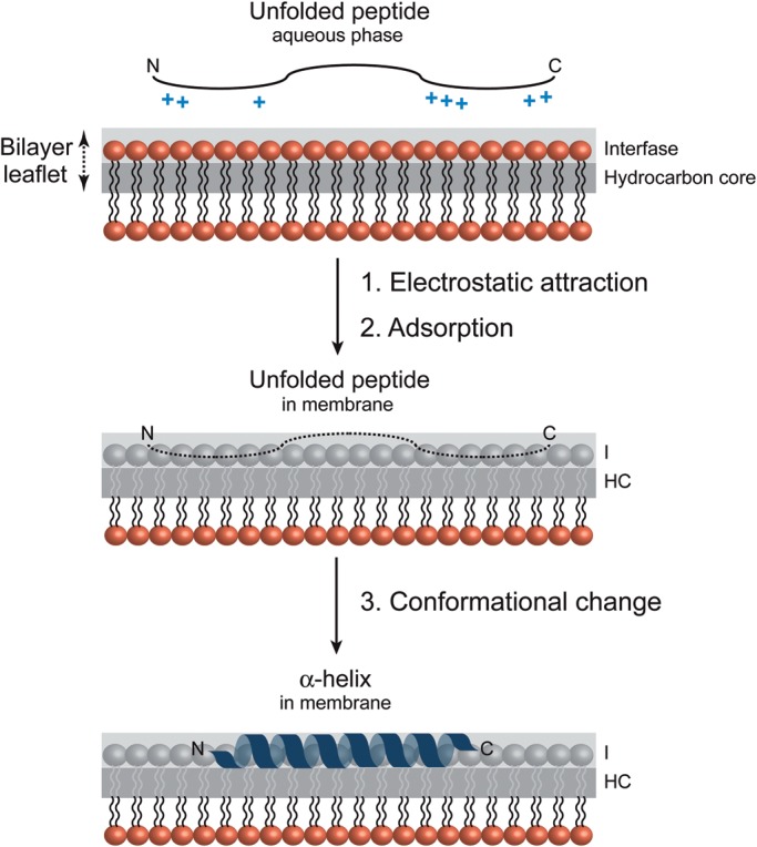
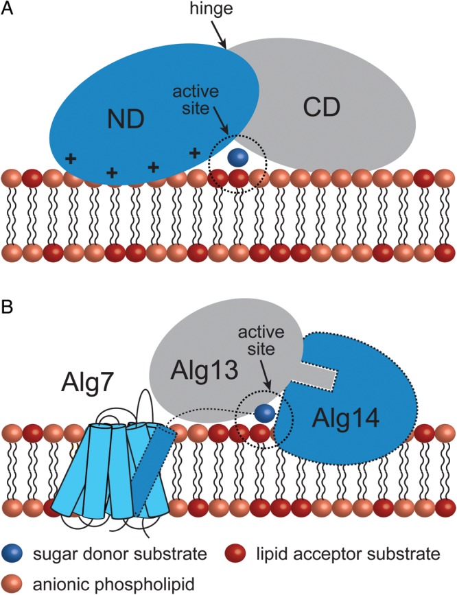
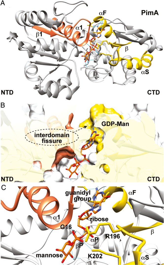
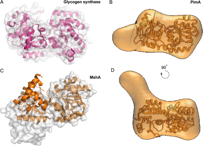
Membrane-associated GT-B glycosyltransferases (GTs) comprise a large family of enzymes that catalyze the transfer of a sugar moiety from nucleotide-sugar donors to a wide range of membrane-associated acceptor substrates, mostly in the form of lipids and proteins. As a consequence, they generate a significant and diverse amount of glycoconjugates in biological membranes, which are particularly important in cell-cell, cell-matrix and host-pathogen recognition events. Membrane-associated GT-B enzymes display two "Rossmann-fold" domains separated by a deep cleft that includes the catalytic center. They associate permanently or temporarily to the phospholipid bilayer by a combination of hydrophobic and electrostatic interactions. They have the remarkable property to access both hydrophobic and hydrophilic substrates that reside within chemically distinct environments catalyzing their enzymatic transformations in an efficient manner. Here, we discuss the considerable progress that has been made in recent years in understanding the molecular mechanism that governs substrate and membrane recognition, and the impact of the conformational transitions undergone by these GTs during the catalytic cycle.

Keywords: X-ray crystallography; carbohydrate-modifying enzyme; glycosyltransferase; membrane protein; structural biology.


Figures

Fig. 1.
Fig. 1.
Catalytic mechanisms in GTs. (A) GTs catalyze the transfer of sugars with either “inversion” or “retention” of the anomeric configuration with respect to the sugar donor substrates. (B) Inverting GTs utilize a direct-displacement SN2-like reaction mechanism involving a single oxocarbenium ion-like transition state. (C) Current mechanisms for enzymatic glycosyl transfer with retention of configuration proposed in the literature: The front-face mechanism and the double-displacement mechanism (Rojas-Cervellera et al. 2013).
Fig. 2.
Fig. 2.
Structural folds in GTs. (A) The overall architecture of the GT-A fold as observed in the dimeric glucosyl 3-phosphoglycerate synthase from M. tuberculosis. The N- and C-terminal domains are shown in orange and yellow, respectively. The second monomer is shown in gray (Urresti et al. 2012). (B) The GT-B fold as visualized in the phosphatidyl-myo-inositol mannosyltransferase PimA from Mycobacterium smegmatis. The N- and C-terminal domains are shown in yellow and orange, respectively (Guerin et al. 2007). (C) GT-C fold members are predicted to have 8–13 transmembrane α-helices with the active site located at the interface between the transmembrane (yellow) and soluble (orange) domains as observed in PglB from Campylobacter lari (Lizak et al. 2011). (D) The peptidoglycan PBP2 from Staphylococcus aureus adopts a fold distinct from those of other GT classes (Lovering et al. 2007).
Fig. 3.
Fig. 3.
Membrane-associated GT-B GTs. (A) A GT-B peripheral membrane GT as illustrated by the phosphatidyl-myo-inositol mannosyltransferase PimA (PM, GT4; Guerin et al. 2007; Guerin et al. 2009). (B) A GT-B monotopic GT as illustrated by the UDP-N-acetylglucosamine–N-acetylmuramyl-(pentapeptide) pyrophosphoryl-undecaprenol N-acetylglucosamine transferase MurG (MT, GT28; Hu et al. 2003) (C) A GT-B bitopic GT as illustrated by the multifunctional sialyltransferase PmST1 from Pasteurella multocida (Ni et al. 2006; Ni et al. 2007). (D) The GT-B dimeric monotopic α-2,3-sialyltransferase NST from Neisseria meningitidis (Gilbert et al. 1997; Lin et al. 2011).
Fig. 4.
Fig. 4.
A model for membrane association of monotopic GT-B GTs. Wieslander and colleagues proposed two modes of reorientation for AtDGD2 upon stimulation by anionic lipids: An up-down displacement (“dipping”) of the C-terminal domain (A) and a “rolling” transfer of the catalytic region into the interface (B). Both models contemplate the access of the soluble UDP-Gal donor substrate to the active site.
Fig. 5.
Fig. 5.
Molecular recognition of a peptide at the membrane surface. Schematic representation of different steps in the process of peptide binding to an anionic phospholipid bilayer. The positively charged peptide is attracted electrostatically to the membrane surface followed by a conformational change to an α-helix (White and Wimley 1999; Seelig 2004).
Fig. 6.
Fig. 6.
Electrostatic surface potential representation of membrane-associated GT-B GTs, as visualized in GumK, MurG, WaaA, WaaC and PimA.
Fig. 7.
Fig. 7.
Calculation of the desolvation energy values derived from octanol/water experiments. The hotspots indicate areas with the most favorable energy change upon binding, likely to be buried in the membrane. The largest hotspots correspond to energy values lower than −15.0 kcal mol−1, medium-sized spots represent values between −15 and −10 kcal mol−1 and the smallest points represent values higher than −10 kcal mol−1.
Fig. 8.
Fig. 8.
Peripheral GT-B GTs adopt different strategies for membrane association. (A) The mycobacterial mannosyltransferase PimA interacts with the phospholipid bilayer by a combination of positively charged and hydrophobic residues exposed on the N-terminal domain of the protein. An important conformational change is expected to occur during/after protein–membrane interaction (Guerin et al. 2007; Guerin et al. 2009; Giganti et al. 2013). (B) A model for the Alg7/13/14 complex formation. This functional multienzyme complex catalyzes the first two steps of lipid-linked oligosaccharide on the cytosolic face of the ER membrane. Alg7, a politopic protein, which transfers a GlcNAc-phosphate to dolichol phosphate, interacts with the transmembrane α-helix of the bitopic GT-GT Alg14 (Lu et al. 2012). Biochemical and structural data support a model in which Alg13-Ag14 interaction is mediated by residues located on the C-terminal α-helix of Alg13 and C-terminal amino acids of Alg14 to form a dimeric Alg13/14 (Wang et al. 2008).
Fig. 9.
Fig. 9.
The nucleotide sugar donor-binding site. (A) Overall structure of the phosphatidylinositol mannosyltransferase PimA (PM, GT4) from M. smegmatis showing the β11 motif (residues Met1 to Ala31) in orange and the α/β/α motif (αF/β/αS; residues Asp252 to Gly287) in yellow (Guerin et al. 2007). (B) Surface representation of the active site of PimA showing the hydrophobic binding pocket and Asp253 that account for nucleoside heterocycle specificity (Guerin et al. 2007). (C) The negatively charged pyrophosphate group is stabilized by α1 (Gly15 to Ala31 at the N-terminal domain) and αS (Gly277 to Gly287 at the C-terminal domain) through helix dipole effect (Aqvist et al. 1991; Hol et al. 1978; Hu and Walker 2002). The α- and β-phosphates are further stabilized by two positively charged residues, Arg196 and Lys202, and the α11 loop.
Fig. 10.
Fig. 10.
Conformational changes on GT-B GTs. (A) Structural comparison between the “open” (molecular surface representation) and “closed” (schematic cartoon) states of glycogen synthase (Sheng et al. 2009) and (C) MshA from C. glutamicum (Vetting et al. 2008). (B, D) Average low-resolution structure of PimA-GDP complex with the high-resolution crystal structure of PimA-GDP complex fitted by rigid body docking (Giganti et al. 2013).

Similar articles

Cited by

References

    1. Abdian PL, Lellouch AC, Gautier C, Ielpi L, Geremia RA. Identification of essential amino acids in the bacterial alpha-mannosyltransferase AceA. J Biol Chem. 2000;275:40568–40575. doi:10.1074/jbc.M007496200. - DOI - PubMed
    1. Andersen OS, Koeppe RE., II Bilayer thickness and membrane protein function: An energetic perspective. Annu Rev Biophys Biomol Struct. 2007;36:107–130. doi:10.1146/annurev.biophys.36.040306.132643. - DOI - PubMed
    1. Andersson AS, Rilfors L, Bergqvist M, Persson S, Lindblom G. New aspects on membrane lipid regulation in Acholeplasma laidlawii A and phase equilibria of monoacyl-diglucosyldiacylglycerol. Biochemistry. 1996;35:11119–11130. doi:10.1021/bi960561w. - DOI - PubMed
    1. Andrés E, Biarnés X, Faijes M, Planas A. Bacterial glycoglycerolipid synthases: Processive and non-processive glycosyltransferases in mycoplasma. Biocatal Biotransfor. 2012;30:274–287. doi:10.3109/10242422.2012.674733. - DOI
    1. Ardévol A, Rovira C. The molecular mechanism of enzymatic glycosyl transfer with retention of configuration: Evidence for a short-lived oxocarbenium-like species. Angew Chem Int Ed Engl. 2011;50:10897–10901. doi:10.1002/anie.201104623. - DOI - PubMed

Publication types