Adult neurogenesis in the mammalian hippocampus: why the dentate gyrus?
- PMID: 24255101
- PMCID: PMC3834622
- DOI: 10.1101/lm.026542.112
Adult neurogenesis in the mammalian hippocampus: why the dentate gyrus?
Abstract
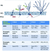
In the adult mammalian brain, newly generated neurons are continuously incorporated into two networks: interneurons born in the subventricular zone migrate to the olfactory bulb, whereas the dentate gyrus (DG) of the hippocampus integrates locally born principal neurons. That the rest of the mammalian brain loses significant neurogenic capacity after the perinatal period suggests that unique aspects of the structure and function of DG and olfactory bulb circuits allow them to benefit from the adult generation of neurons. In this review, we consider the distinctive features of the DG that may account for it being able to profit from this singular form of neural plasticity. Approaches to the problem of neurogenesis are grouped as "bottom-up," where the phenotype of adult-born granule cells is contrasted to that of mature developmentally born granule cells, and "top-down," where the impact of altering the amount of neurogenesis on behavior is examined. We end by considering the primary implications of these two approaches and future directions.
Figures


Similar articles
-
Continuous postnatal neurogenesis contributes to formation of the olfactory bulb neural circuits and flexible olfactory associative learning.J Neurosci. 2014 Apr 23;34(17):5788-99. doi: 10.1523/JNEUROSCI.0674-14.2014. J Neurosci. 2014. PMID: 24760839 Free PMC article.
-
Adult neurogenesis modifies excitability of the dentate gyrus.Front Neural Circuits. 2013 Dec 26;7:204. doi: 10.3389/fncir.2013.00204. eCollection 2013. Front Neural Circuits. 2013. PMID: 24421758 Free PMC article.
-
Early survival and delayed death of developmentally-born dentate gyrus neurons.Hippocampus. 2017 Nov;27(11):1155-1167. doi: 10.1002/hipo.22760. Epub 2017 Jul 18. Hippocampus. 2017. PMID: 28686814
-
Adult neurogenesis in the mammalian dentate gyrus.Anat Histol Embryol. 2020 Jan;49(1):3-16. doi: 10.1111/ahe.12496. Epub 2019 Sep 30. Anat Histol Embryol. 2020. PMID: 31568602 Review.
-
Contributions of adult neurogenesis to dentate gyrus network activity and computations.Behav Brain Res. 2019 Nov 18;374:112112. doi: 10.1016/j.bbr.2019.112112. Epub 2019 Aug 1. Behav Brain Res. 2019. PMID: 31377252 Free PMC article. Review.
Cited by
-
Bidirectional communication between the innate immune and nervous systems for homeostatic neurogenesis in the adult hippocampus.Neurogenesis (Austin). 2015 Nov 25;2(1):e1081714. doi: 10.1080/23262133.2015.1081714. eCollection 2015. Neurogenesis (Austin). 2015. PMID: 27604264 Free PMC article.
-
Reliable Genetic Labeling of Adult-Born Dentate Granule Cells Using Ascl1 CreERT2 and Glast CreERT2 Murine Lines.J Neurosci. 2015 Nov 18;35(46):15379-90. doi: 10.1523/JNEUROSCI.2345-15.2015. J Neurosci. 2015. PMID: 26586824 Free PMC article.
-
How Do Microtubule Dynamics Relate to the Hallmarks of Learning and Memory?J Neurosci. 2016 Jun 1;36(22):5911-3. doi: 10.1523/JNEUROSCI.0920-16.2016. J Neurosci. 2016. PMID: 27251613 Free PMC article. Review. No abstract available.
-
Cognitive Reserve and the Prevention of Dementia: the Role of Physical and Cognitive Activities.Curr Psychiatry Rep. 2016 Sep;18(9):85. doi: 10.1007/s11920-016-0721-2. Curr Psychiatry Rep. 2016. PMID: 27481112 Free PMC article. Review.
-
The Possible Roles of the Dentate Granule Cell's Leptin and Other Ciliary Receptors in Alzheimer's Neuropathology.Cells. 2015 Jul 13;4(3):253-74. doi: 10.3390/cells4030253. Cells. 2015. PMID: 26184316 Free PMC article. Review.
References
-
- Adams B, Lee M, Fahnestock M, Racine RJ 1997. Long-term potentiation trains induce mossy fiber sprouting. Brain Res 775: 193–197 - PubMed
-
- Aimone JB, Wiles J, Gage FH 2006. Potential role for adult neurogenesis in the encoding of time in new memories. Nat Neurosci 9: 723–727 - PubMed
-
- Airan RD, Meltzer LA, Roy M, Gong Y, Chen H, Deisseroth K 2007. High-speed imaging reveals neurophysiological links to behavior in an animal model of depression. Science 317: 819–823 - PubMed
Publication types
MeSH terms
Grants and funding
LinkOut - more resources
Full Text Sources
Other Literature Sources
