. 2013 Nov 19;4(6):e00721-13.
doi: 10.1128/mBio.00721-13.

cor, a novel carbon monoxide resistance gene, is essential for Mycobacterium tuberculosis pathogenesis

Affiliations

cor, a novel carbon monoxide resistance gene, is essential for Mycobacterium tuberculosis pathogenesis

Vineetha M Zacharia et al. mBio. .

Abstract

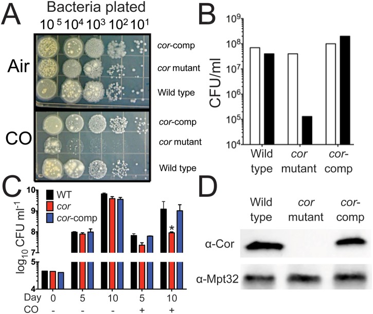
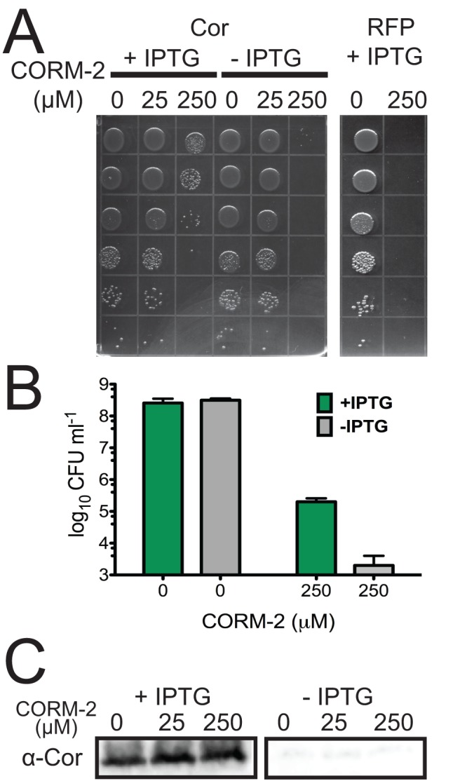
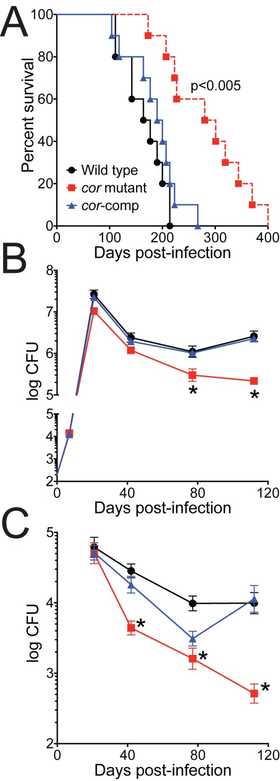
Tuberculosis, caused by Mycobacterium tuberculosis, remains a devastating human infectious disease, causing two million deaths annually. We previously demonstrated that M. tuberculosis induces an enzyme, heme oxygenase (HO1), that produces carbon monoxide (CO) gas and that M. tuberculosis adapts its transcriptome during CO exposure. We now demonstrate that M. tuberculosis carries a novel resistance gene to combat CO toxicity. We screened an M. tuberculosis transposon library for CO-susceptible mutants and found that disruption of Rv1829 (carbon monoxide resistance, Cor) leads to marked CO sensitivity. Heterologous expression of Cor in Escherichia coli rescued it from CO toxicity. Importantly, the virulence of the cor mutant is attenuated in a mouse model of tuberculosis. Thus, Cor is necessary and sufficient to protect bacteria from host-derived CO. Taken together, this represents the first report of a role for HO1-derived CO in controlling infection of an intracellular pathogen and the first identification of a CO resistance gene in a pathogenic organism.

Importance: Macrophages produce a variety of antimicrobial molecules, including nitric oxide (NO), hydrogen peroxide (H2O2), and acid (H+), that serve to kill engulfed bacteria. In addition to these molecules, human and mouse macrophages also produce carbon monoxide (CO) gas by the heme oxygenase (HO1) enzyme. We observed that, in contrast to other bacteria, mycobacteria are resistant to CO, suggesting that this might be an evolutionary adaptation of mycobacteria for survival within macrophages. We screened a panel of ~2,500 M. tuberculosis mutants to determine which genes are required for survival of M. tuberculosis in the presence of CO. Within this panel, we identified one such gene, cor, that specifically confers CO resistance. Importantly, we found that the ability of M. tuberculosis cells carrying a mutated copy of this gene to cause tuberculosis in a mouse disease model is significantly attenuated. This indicates that CO resistance is essential for mycobacterial survival in vivo.


Figures

FIG 1
FIG 1
Identification of cor (Rv1829) as a CO resistance gene. (A) Serial dilutions of M. tuberculosis Erdman, cor mutant bacteria, or the cor mutant complemented with cor (cor-comp) were plated and exposed to ambient air or CO (0.2%) for 3 weeks. (B) Quantitation of CFU data from the experiment represented by panel A (one of three similar experiments shown). (C) Western blots of lysates of M. tuberculosis Erdman, the cor mutant, or the complemented strain were probed with anti-Cor antibody. WT, wild type. (D) M. tuberculosis Erdman, the cor mutant, and the complemented strain were grown in 7H9 liquid medium in the presence or absence of CO, and CFU were enumerated. *, P < 0.05 compared to M. tuberculosis Erdman (by Student’s t test).
FIG 2
FIG 2
Cor is a conserved, ancient protein. (A) Alignment of Rv1829 from M. tuberculosis to orthologues from Thermotoga maritima, M. leprae, M. smegmatis, Rhodococcus fascians, and Streptomyces species AA4. Sequence alignment of Rv1829 homologues shows conserved amino acids (in rainbow colors) by conservation, with invariant nonhydrophobic residues colored in red. This color scheme is identical in the modeled crystal structure in panel D. (B) Species-distribution taxonomic tree in “sunburst” format. The distribution and evolutionary conservation of all known homologues of Cor are shown. (C) Alignment of the Cor genomic region from M. tuberculosis with the orthologous genomic region from Rhodococcus sp. demonstrates conservation of multiple surrounding genes. (D) The Cor sequence was mapped to the representative DUF151 structure, which highlighted a conserved surface cleft formed at the interface (also relatively conserved) of two DUF151 monomers (shown in red). (E) Cor was purified from E. coli as a 6×His-tagged protein and then run on a denaturing SDS-PAGE gel (left panel) or a native gel (right panel) with appropriate molecular mass markers. The predicted molecular masses of cor are 18 kDa for the monomer and 36 kDa for the dimer. In the left panel, lanes are total lysate (lane 1), flowthrough (lane 2), final wash (lane 3), and imidazole elution (lane 4).
FIG 3
FIG 3
The cor mutant is not hypersusceptible to acid pH, nitric oxide, hypoxia, or hydrogen peroxide. (A) M. tuberculosis Erdman, the cor mutant, and the cor mutant complemented with cor were diluted into 7H9-ADN to an OD600 of 0.05 at pH 5.5 or pH 5.5 plus sodium nitrite. Six days later, bacteria were diluted and plated and CFU enumerated after 3 weeks. (B) M. tuberculosis Erdman, the cor mutant, and the complemented strain (cor-comp) were grown to late log phase and resuspended to an OD600 of 0.1, and bacteria were plated to generate a dense lawn. Sterile paper discs placed on the plate were impregnated with 10 µl plumbagin, plates were incubated for 3 weeks, and the zone of inhibition was measured. (C and D) M. tuberculosis Erdman (black circles) and the cor mutant (red squares) were grown in 17-ml test tubes in triplicate, and gradual hypoxia was generated following the Wayne model. At each time point, tubes were sacrificed and OD600 (C) and CFU (D) were determined. Data represent the results of one of three similar experiments. Data represent means ± standard deviations (SD) for both panels.
FIG 4
FIG 4
The cor mutant has a dysregulated intracellular redox environment, and its CO susceptibility can be rescued by the antioxidant α-tocopherol. (A and B) M. tuberculosis cultures were exposed to CO in vitro in quadruplicate and harvested after 0, 5, and 10 days. Small-molecule metabolites were extracted, and 145 known biochemicals were quantified using GC-MS and LC-MS. Normalized metabolite levels produced under the indicated treatment conditions, where the mean metabolite value for wild-type M. tuberculosis served as the denominator, are shown. Compared to wild-type bacteria, the cor mutant treated with CO had significantly reduced levels of mycothione (A) and NAD+ (B) but had increased levels of linoleate (C), linolenate (D), palmitoleate (E), and nonadecanoate (F). Differences between CO-treated M. tuberculosis Erdman and the mutant at 10 days were statistically significant. *, P < 0.05 (by Welch’s two-sample t test).
FIG 5
FIG 5
E. coli CO susceptibility is prevented by cor expression. (A) E. coli transformed with an IPTG-inducible vector expressing Cor or RFP was exposed to the CO donor corm-2 for 30 min with or without prior IPTG treatment, and serial dilutions were plated. (B) E. coli was grown as described above, and serial dilutions were plated to determine surviving CFU. (C) E. coli expressing Cor was lysed and Cor accumulation determined by Western blot analysis.
FIG 6
FIG 6
Virulence of the cor mutant is attenuated in mice. BALBc mice were infected with 102 CFU wild-type (WT) M. tuberculosis, the cor mutant, or the cor mutant complemented with cor via aerosol. (A) Ten mice per group were monitored over time for survival. By Kaplan-Meier analysis, differences between groups infected with the cor mutant and M. tuberculosis Erdman or the complemented strain are statistically significant. (B and C) Five mice per group were sacrificed, mouse lung (B) and mouse liver (C) were harvested and homogenized, and CFU were enumerated. *, P < 0.05 compared to WT (by the nonparametric Kruskal-Wallis test).

References

    1. Huynh KK, Joshi SA, Brown EJ. 2011. A delicate dance: host response to mycobacteria. Curr. Opin. Immunol. 23:464–472 - PubMed
    1. Liu PT, Modlin RL. 2008. Human macrophage host defense against Mycobacterium tuberculosis. Curr. Opin. Immunol. 20:371–376 - PubMed
    1. Nathan C, Shiloh MU. 2000. Reactive oxygen and nitrogen intermediates in the relationship between mammalian hosts and microbial pathogens. Proc. Natl. Acad. Sci. U. S. A. 97:8841–8848 - PMC - PubMed
    1. Rohde K, Yates RM, Purdy GE, Russell DG. 2007. Mycobacterium tuberculosis and the environment within the phagosome. Immunol. Rev. 219:37–54 - PubMed
    1. Davis AS, Vergne I, Master SS, Kyei GB, Chua J, Deretic V. 2007. Mechanism of inducible nitric oxide synthase exclusion from mycobacterial phagosomes. PLOS Pathog. 3:e186.10.1371/journal.ppat.0030186 - DOI - PMC - PubMed

Publication types

MeSH terms

LinkOut - more resources