

Use of (18)F-FDG PET/CT to locate primary malignancies in patients with hepatic cirrhosis and malignant ascites
- PMID: 24255572
- PMCID: PMC3828429
- DOI: 10.3978/j.issn.1000-9604.2013.09.01
Use of (18)F-FDG PET/CT to locate primary malignancies in patients with hepatic cirrhosis and malignant ascites
Abstract
Objective: Ascites in patients with hepatic cirrhosis is caused by cirrhosis in most cases. For most malignant ascites, the primary malignancy could be readily identified using conventional imaging methods, e.g., computed tomography (CT) and magnetic resonance imaging (MRI). However, in a small fraction of the patients, the primary malignancy remains occult even with these examinations. In this retrospective study, we assessed the usefulness of (18)F-FDG PET/CT in patients with hepatic cirrhosis and malignant ascites of otherwise unknown origin.
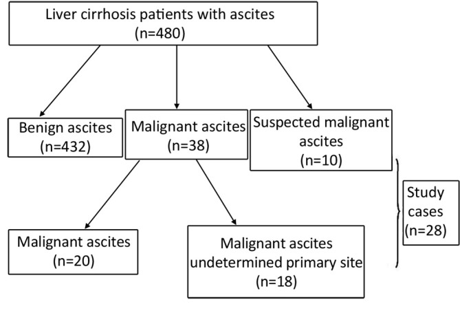
Methods: Twenty-eight patients with malignant ascites of unknown primary sites after CT, MRI and ultrasound during the period of five years between January 2008 and December 2012 had received (18)F-FDG PET/CT. Medical records of these patients were reviewed and analyzed.
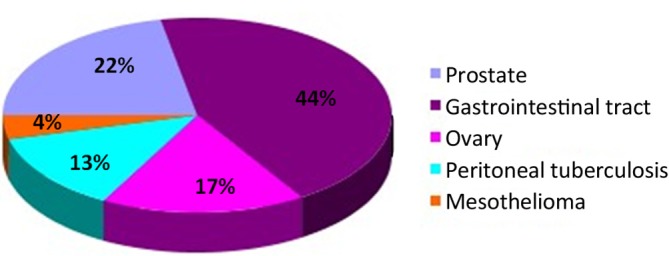
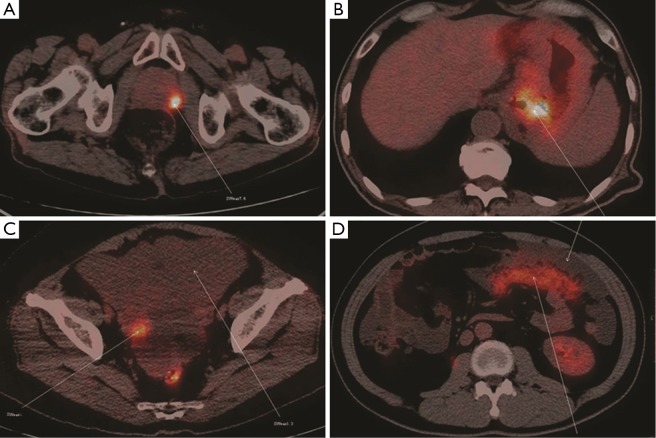
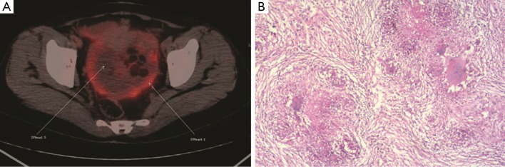
Results: Elevated (18)F-FDG absorption was found in 23 of 28 cases in the following sites: gastrointestinal tract (n=10, 43.5%), prostate (n=5, 21.7%), peritoneum (n=4, 13.3%), and ovary (n=4, 13.3%). Cancer was confirmed by pathology in 20 cases after open or laparoscopic surgeries. Five patients were found to have benign ascites, among which, 3 were found to be false positive due to tuberculosis. SUV values were significantly higher for tumors than for benign lesions (mean values, 6.95 vs. 2.94; P=0.005).
Conclusions: The (18)F-FDG PET/CT can be as a powerful imaging tool in identifying tissue origin in liver cirrhosis patients suspected of cancers or with cancers of unknown primary sites.
Keywords: PET/CT; cancer, ascites; liver cirrhosis.
Figures
Similar articles
-
More advantages in detecting bone and soft tissue metastases from prostate cancer using 18F-PSMA PET/CT.Hell J Nucl Med. 2019 Jan-Apr;22(1):6-9. doi: 10.1967/s002449910952. Epub 2019 Mar 7. Hell J Nucl Med. 2019. PMID: 30843003
-
The Role of 18F-FDG PET/CT in the evaluation of Ascites of Undetermined Origin.J Nucl Med. 2009 Apr;50(4):506-12. doi: 10.2967/jnumed.108.056382. Epub 2009 Mar 16. J Nucl Med. 2009. PMID: 19289438
-
Clinical value of 18F-FDG-PET/CT in staging cutaneous squamous cell carcinoma.Nucl Med Commun. 2019 Jul;40(7):744-751. doi: 10.1097/MNM.0000000000001029. Nucl Med Commun. 2019. PMID: 31095044 Free PMC article.
-
Indirect comparison of the diagnostic performance of 18F-FDG PET/CT and MRI in differentiating benign and malignant ovarian or adnexal tumors: a systematic review and meta-analysis.BMC Cancer. 2021 Oct 6;21(1):1080. doi: 10.1186/s12885-021-08815-3. BMC Cancer. 2021. PMID: 34615498 Free PMC article.
-
Dual-time point 18F-FDG-PET and PET/CT for Differentiating Benign From Malignant Musculoskeletal Lesions: Opportunities and Limitations.Semin Nucl Med. 2017 Jul;47(4):373-391. doi: 10.1053/j.semnuclmed.2017.02.009. Epub 2017 Apr 19. Semin Nucl Med. 2017. PMID: 28583277 Review.
References
-
- Reynolds TB. Ascites. Clin Liver Dis 2000;4:151-68 - PubMed
-
- European Association for the Study of the Liver EASL clinical practice guidelines on the management of ascites, spontaneous bacterial peritonitis, and hepatorenal syndrome in cirrhosis. J Hepatol 2010;53:397-417 - PubMed
-
- Moore KP, Wong F, Gines P, et al. The management of ascites in cirrhosis: report on the consensus conference of the International Ascites Club. Hepatology 2003;38:258-66 - PubMed
-
- Han CM, Lee CL, Huang KG, et al. Diagnostic laparoscopy in ascites of unknown origin: Chang Gung Memorial Hospital 20-year experience. Chang Gung Med J 2008;31:378-83 - PubMed
-
- Li XJ, Li FQ, Han JK, et al. Ascites metabolism measurement enhanced the diagnostic value and accuracy of prognostic evaluation in 18F-FDG PET/CT studies in malignant ascites patients. Nucl Med Commun 2013;34:544-50 - PubMed
LinkOut - more resources
Full Text Sources