Morphologic alteration of metastatic neuroblastic tumor in bone marrow after chemotherapy
- PMID: 24255631
- PMCID: PMC3830990
- DOI: 10.4132/KoreanJPathol.2013.47.5.433
Morphologic alteration of metastatic neuroblastic tumor in bone marrow after chemotherapy
Abstract
Background: The aim of this study is to evaluate the histologic features of metastatic neuroblastic tumors (NTs) in bone marrow (BM) before and after chemotherapy in comparison with those of primary NTs.
Methods: A total of 294 biopsies from 48 children diagnosed with NTs with BM metastasis were examined. There were 48 primary neoplasm biopsies, 48 BM biopsies before chemotherapy, 36 primary neoplasm excisional biopsies after chemotherapy, and 162 BM biopsies after chemotherapy.
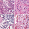
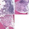
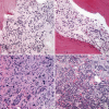
Results: Metastatic NTs in BM before chemotherapy were composed of undifferentiated and/or differentiating neuroblasts, but had neither ganglion cells nor Schwannian stroma. Metastatic foci of BM after chemotherapy were found to have differentiated into ganglion cells or Schwannian stroma, which became more prominent after further cycles of chemotherapy. Persistence of NTs or tumor cell types in BM after treatment did not show statistically significant correlation to patients' outcome. However, three out of five patients who newly developed poorly differentiated neuroblasts in BM after treatment expired due to disease progression.
Conclusions: Metastatic NTs in BM initially consist of undifferentiated or differentiating neuroblasts regardless of the primary tumor subtype, and become differentiated after chemotherapy. Newly appearing poorly differentiated neuroblasts after treatment might be an indicator for poor prognosis.
Keywords: Bone marrow; Drug therapy; Histology; Neoplasm metastasis; Neuroblastoma.
Conflict of interest statement
No potential conflict of interest relevant to this article was reported.
Figures


References
-
- Esiashvili N, Goodman M, Ward K, Marcus RB, Jr, Johnstone PA. Neuroblastoma in adults: incidence and survival analysis based on SEER data. Pediatr Blood Cancer. 2007;49:41–46. - PubMed
-
- Bernstein ML, Leclerc JM, Bunin G, et al. A population-based study of neuroblastoma incidence, survival, and mortality in North America. J Clin Oncol. 1992;10:323–329. - PubMed
-
- Moon SB, Park KW, Jung SE, Youn WJ. Neuroblastoma: treatment outcome after incomplete resection of primary tumors. Pediatr Surg Int. 2009;25:789–793. - PubMed
-
- Jung G, Kim J. Primary hepatic neuroblastoma: a case report. Korean J Pathol. 2011;45:423–427.
-
- Moss TJ, Reynolds CP, Sather HN, Romansky SG, Hammond GD, Seeger RC. Prognostic value of immunocytologic detection of bone marrow metastases in neuroblastoma. N Engl J Med. 1991;324:219–226. - PubMed
LinkOut - more resources
Full Text Sources
Other Literature Sources

