Fine needle aspiration cytology of parathyroid lesions
- PMID: 24255635
- PMCID: PMC3830994
- DOI: 10.4132/KoreanJPathol.2013.47.5.466
Fine needle aspiration cytology of parathyroid lesions
Abstract
Background: There has been an increase in the use of fine needle aspiration cytology (FNAC) for the diagnosis of parathyroid lesions (PLs). Differentiation between a thyroid lesion and a PL is not easy because of their similar features. We reviewed parathyroid aspirates in our institution and aimed to uncover trends in diagnostic criteria.
Methods: We selected 25 parathyroid aspirates (from 6 men and 19 women) confirmed surgically or immunohistochemically from 2006 to 2011.
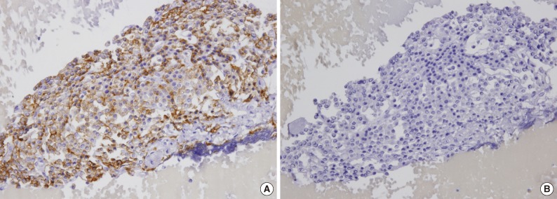
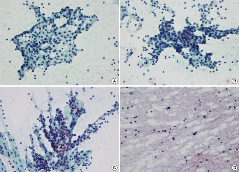
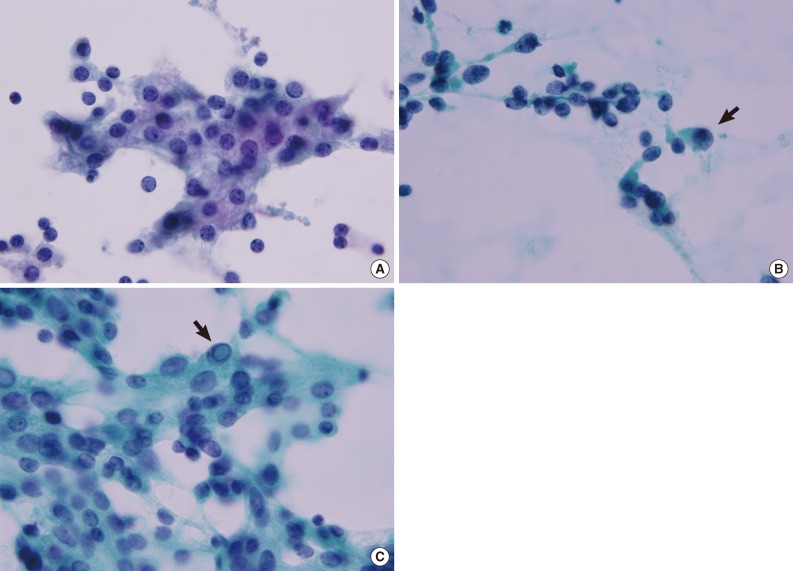
Results: Major architectural findings of PLs include scattered naked nuclei, loose clusters, a papillary pattern with a fibrovascular core, tight clusters, and a follicular pattern. These architectures were commonly admixed with one another. Cytological features included anisokaryosis, stippled chromatin, a well-defined cell border, and oxyphilic cytoplasm. Eighteen of the 25 patients were diagnosed with PL using FNAC. Seven patients had been misdiagnosed with atypical cells (n=2), benign follicular cells (n=2), adenomatous goiter (n=2) and metastatic carcinoma (n=1) in FNAC. Using clinicoradiologic data, the sensitivity of the cytological diagnosis was 86.7%. The cytological sensitivity decreased to 50% without this information.
Conclusions: FNAC of PL is easily confused with thyroid lesions. A combination of cytological parameters and clinical data will be required to improve the diagnostic sensitivity of PLs.
Keywords: Biopsy, fine-needle; Cytology; Parathyroid lesions.
Conflict of interest statement
No potential conflict of interest relevant to this article was reported.
Figures
References
-
- Liu F, Gnepp DR, Pisharodi LR. Fine needle aspiration of parathyroid lesions. Acta Cytol. 2004;48:133–136. - PubMed
-
- Absher KJ, Truong LD, Khurana KK, Ramzy I. Parathyroid cytology: avoiding diagnostic pitfalls. Head Neck. 2002;24:157–164. - PubMed
-
- Agarwal AM, Bentz JS, Hungerford R, Abraham D. Parathyroid fine-needle aspiration cytology in the evaluation of parathyroid adenoma: cytologic findings from 53 patients. Diagn Cytopathol. 2009;37:407–410. - PubMed
-
- Solbiati L, Montali G, Croce F, Bellotti E, Giangrande A, Ravetto C. Parathyroid tumors detected by fine-needle aspiration biopsy under ultrasonic guidance. Radiology. 1983;148:793–797. - PubMed
LinkOut - more resources
Full Text Sources
Other Literature Sources