Abdominal fibromatosis in a young child: a case study and review of the literature
- PMID: 24255636
- PMCID: PMC3830995
- DOI: 10.4132/KoreanJPathol.2013.47.5.472
Abdominal fibromatosis in a young child: a case study and review of the literature
Abstract
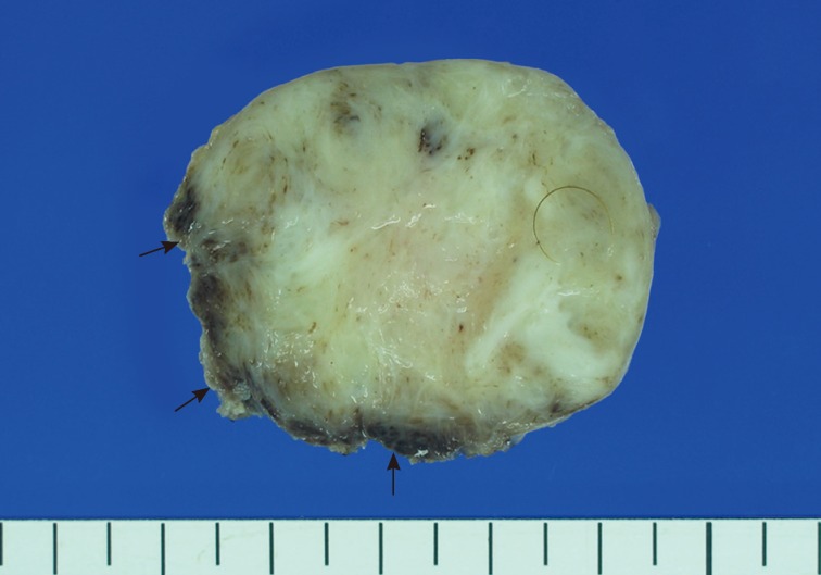
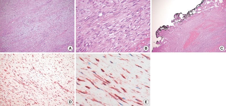
Fibromatoses comprise many different entities of well-differentiated fibroblastic proliferation with variable collagen production and form a firm nodular mass. Abdominal fibromatosis is distinguishable from other forms of fibromatosis because of its location and its tendency to occur in women of childbearing age during or following pregnancy. Abdominal fibromatosis in children is an extremely rare condition. A 15-month-old boy presented with an abdominal wall mass that had recently increased in size. Mass excision was perfomed. The tumor was 4.3×4.1 cm and partly circumscribed. Histologically, the tumor was composed of parallel long fascicles of spindle-cells with a uniform appearance. The edges of the resected mass were infiltrative, and the surgical margins were positive. Mitotic figures were <1/10 high power fields. No cellular atypia or necrosis was present. The tumor cells were positive for vimentin and nuclear β-catenin staining.
Keywords: Abdominal wall; Child; Fibromatosis.
Conflict of interest statement
No potential conflict of interest relevant to this article was reported.
Figures
References
-
- Weiss SW, Goldblum JR. Enzinger and Weiss's soft tissue tumors. Philadelphia: Mosby Elsevier; 2008. pp. 277–302.
-
- Ademuyiwa AO, Bode CO, Elebute OA. Anterior abdominal wall desmoids tumor in a five year old girl: a pre operative diagnostic challenge in resource-poor setting. Ann Pediatr Surg. 2010;6:41–43.
-
- Atahan IL, Akyol F, Zorlu F, Gürkaynak M. Radiotherapy in the management of aggressive fibromatosis. Br J Radiol. 1989;62:854–856. - PubMed
-
- Ayala AG, Ro JY, Goepfert H, Cangir A, Khorsand J, Flake G. Desmoid fibromatosis: a clinicopathologic study of 25 children. Semin Diagn Pathol. 1986;3:138–150. - PubMed
-
- Buitendijk S, van de Ven CP, Dumans TG, et al. Pediatric aggressive fibromatosis: a retrospective analysis of 13 patients and review of literature. Cancer. 2005;104:1090–1099. - PubMed
LinkOut - more resources
Full Text Sources
Other Literature Sources