

A case of unusual visceral heterotaxy syndrome with isolated levocardia
- PMID: 24255657
- PMCID: PMC3831019
- DOI: 10.4070/kcj.2013.43.10.705
A case of unusual visceral heterotaxy syndrome with isolated levocardia
Abstract
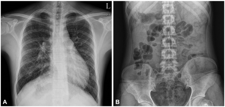
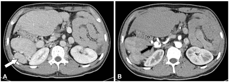
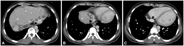
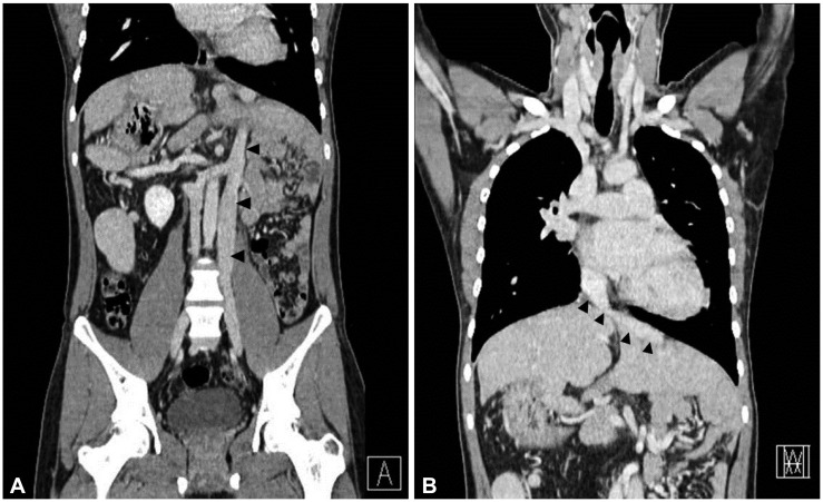
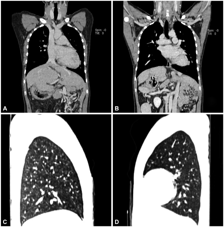
Situs inversus of the abdominal organs in the presence of normally placed heart on the left side of the thorax is known as situs inversus with isolated levocardia. This rare condition is commonly associated with severe congenital defects of the heart. We report a case of situs inversus with levocardia in a 19-year-old asymptomatic male patient with completely normal heart on the left chest. Spiral computed tomography of the thorax and abdomen and echocardiographic studies revealed situs inversus of abdominal organs, normal heart (levocardia), mirrored left lungs, a midline liver, a left-sided inferior vena cava connecting to the right atrium, multiple splenic masses in the abdominal right upper quadrant, and aneurysmal dilatation of a splenic artery.
Keywords: Heterotaxy syndrome; Levocardia.
Conflict of interest statement
The authors have no financial conflicts of interest.
Figures
References
-
- Liberthson RR, Hastreiter AR, Sinha SN, Bharati S, Novak GM, Lev M. Levocardia with visceral heterotaxy--isolated levocardia: pathologic anatomy and its clinical implications. Am Heart J. 1973;85:40–54. - PubMed
-
- Gindes L, Hegesh J, Barkai G, Jacobson JM, Achiron R. Isolated levocardia: prenatal diagnosis, clinical importance, and literature review. J Ultrasound Med. 2007;26:361–365. - PubMed
-
- Moller JH, Nakib A, Anderson RC, Edwards JE. Congenital cardiac disease associated with polysplenia. A developmental complex of bilateral "left-sidedness". Circulation. 1967;36:789–799. - PubMed
-
- Imamura T, Momoi N, Go H, Ogasawara K, Sato M, Hosoya M. Rare case of isolated levocardia with polysplenia including normally structured lung without cardiac anomaly. Congenit Anom (Kyoto) 2011;51:187–190. - PubMed
LinkOut - more resources
Full Text Sources
Other Literature Sources
