. 2013 Nov 8;8(11):e79530.
doi: 10.1371/journal.pone.0079530. eCollection 2013.

β-lactoglobulin's conformational requirements for ligand binding at the calyx and the dimer interphase: a flexible docking study

Affiliations

β-lactoglobulin's conformational requirements for ligand binding at the calyx and the dimer interphase: a flexible docking study

Lenin Domínguez-Ramírez et al. PLoS One. .

Abstract

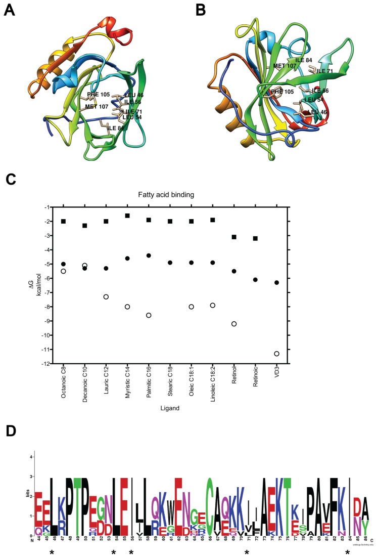
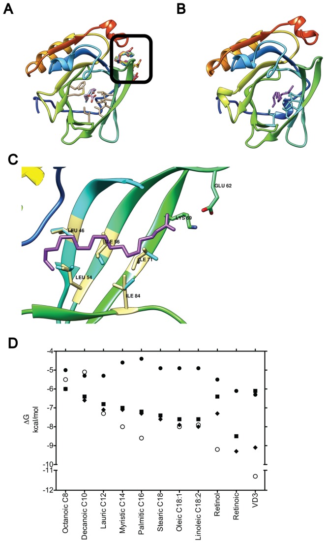
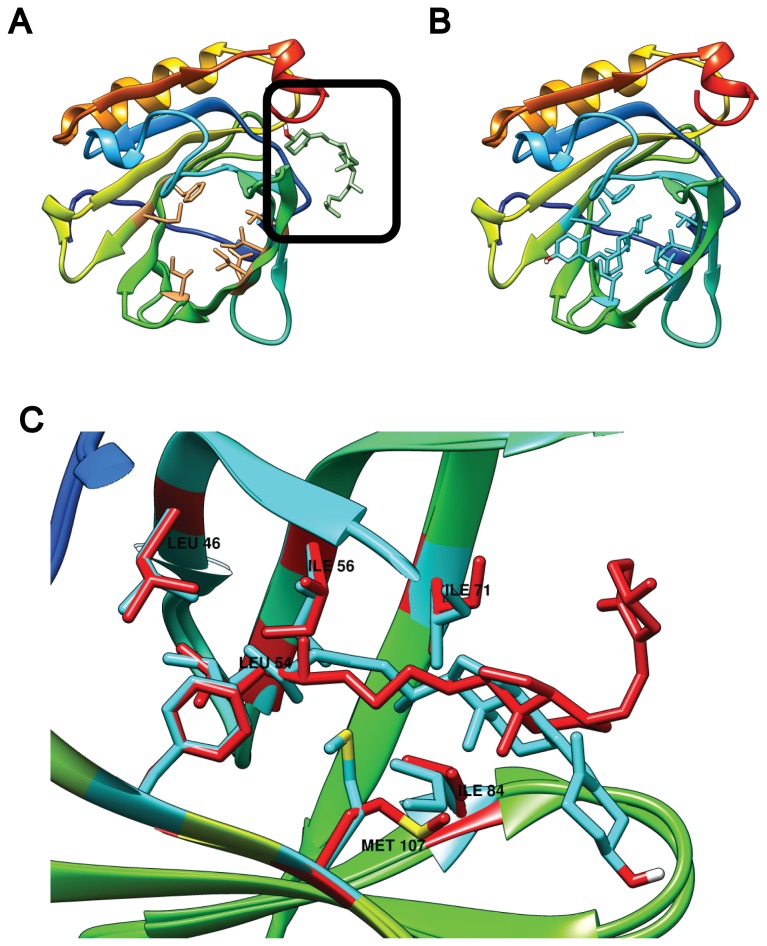
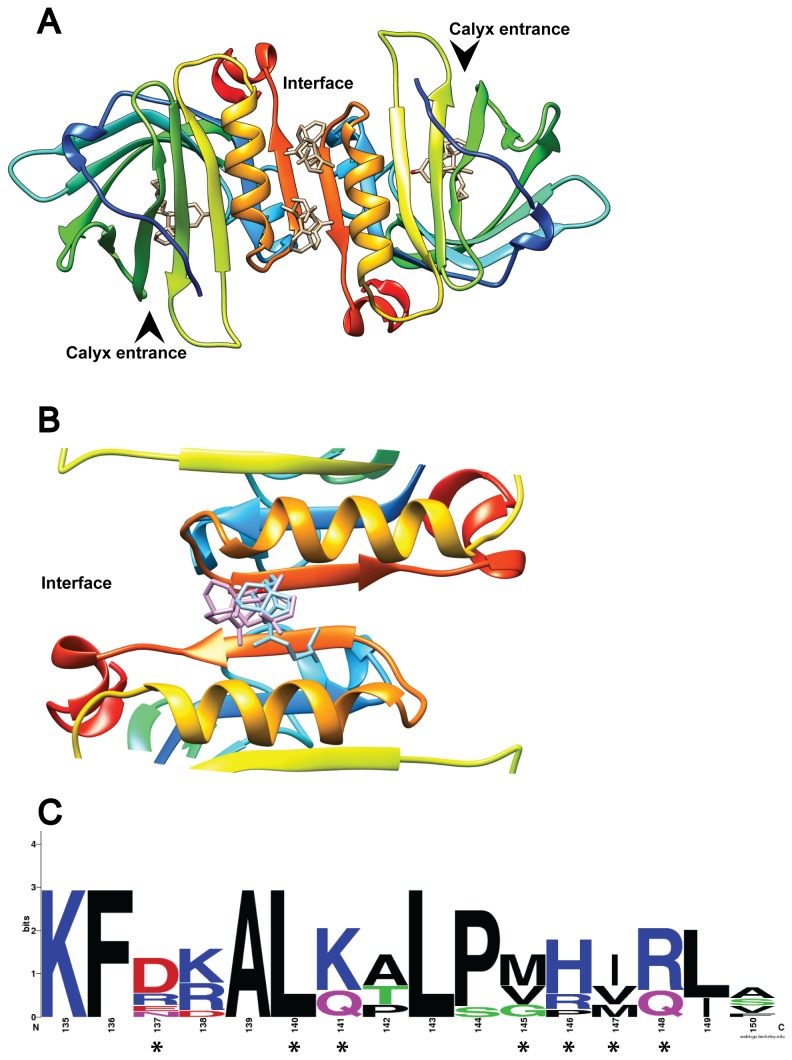
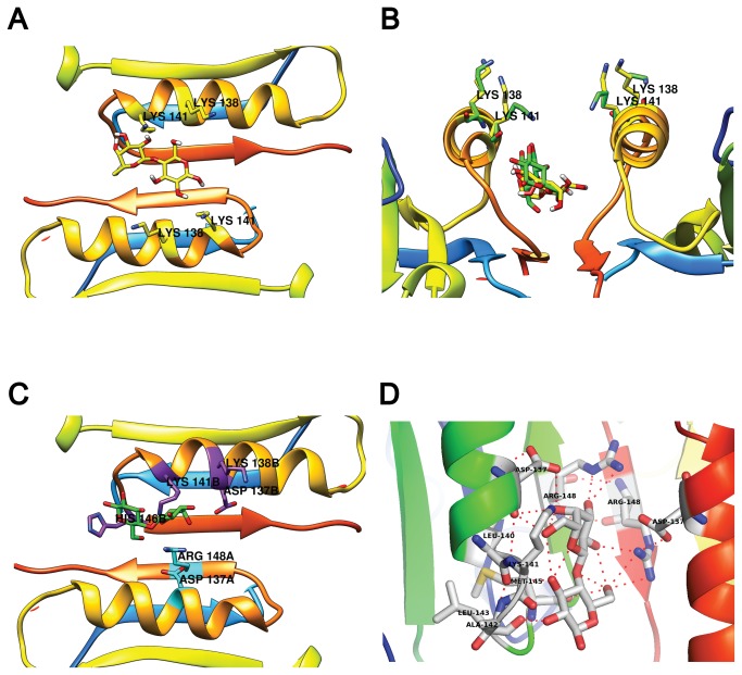
β-lactoglobulin (BLG) is an abundant milk protein relevant for industry and biotechnology, due significantly to its ability to bind a wide range of polar and apolar ligands. While hydrophobic ligand sites are known, sites for hydrophilic ligands such as the prevalent milk sugar, lactose, remain undetermined. Through the use of molecular docking we first, analyzed the known fatty acid binding sites in order to dissect their atomistic determinants and second, predicted the interaction sites for lactose with monomeric and dimeric BLG. We validated our approach against BLG structures co-crystallized with ligands and report a computational setup with a reduced number of flexible residues that is able to reproduce experimental results with high precision. Blind dockings with and without flexible side chains on BLG showed that: i) 13 experimentally-determined ligands fit the calyx requiring minimal movement of up to 7 residues out of the 23 that constitute this binding site. ii) Lactose does not bind the calyx despite conformational flexibility, but binds the dimer interface and an alternate Site C. iii) Results point to a probable lactolation site in the BLG dimer interface, at K141, consistent with previous biochemical findings. In contrast, no accessible lysines are found near Site C. iv) lactose forms hydrogen bonds with residues from both monomers stabilizing the dimer through a claw-like structure. Overall, these results improve our understanding of BLG's binding sites, importantly narrowing down the calyx residues that control ligand binding. Moreover, our results emphasize the importance of the dimer interface as an insufficiently explored, biologically relevant binding site of particular importance for hydrophilic ligands. Furthermore our analyses suggest that BLG is a robust scaffold for multiple ligand-binding, suitable for protein design, and advance our molecular understanding of its ligand sites to a point that allows manipulation to control binding.


Conflict of interest statement

Competing Interests: The authors gratefully acknowledge the support of NVIDIA Corporation with the donation of the Tesla K20 GPU used for this research. However, this donation/relationship with NVIDIA does not alter the authors' adherence to all the PLOS ONE policies on sharing data and materials nor influences the authors' research in any way.

Figures

Figure 1
Figure 1. β-lactoglobulin and its calyx binding site.
The main BLG binding site (calyx or Site A) is shown empty from two perspectives: (A) top-down view and (B) bottom-up view. The seven flexible residues required for ligand binding are labeled. The secondary structure is colored from N-terminus in blue, to C-terminus in red. (C) Plot of binding energy calculated from docking vs ligand using rigid, monomeric, empty BLG structures with open (2BLG, black circles), or semi closed (2Q39, black squares) EF loops, and compared to experimentally determined data (open circles). Fatty acids are sorted by increasing size, or in the case of stearic, oleic and linoleic, by decreasing saturation. No experimental affinity has been reported for stearic or retinoic acids. (D) Weblogo of the sequence alignment of BLG from 7 mammals. Asterisks indicate the 5 residues made flexible for docking.
Figure 2
Figure 2. Effect of residue flexibility on fatty acid binding.
(A) Docking to a rigid 2BLG allows only the three smallest lipids into the calyx (shown superposed in white, light blue and light purple), while excluding longer fatty acids to Site C (black square, fatty acids in different colors). The seven binding residues in the calyx are shown in light yellow. When five of these residues were allowed flexibility all fatty acids bind the calyx. Stearic acid (purple) is shown bound in (B) and (C) in a full BLG top down view and a side view magnification of the calyx, respectively. In (C) the five flexible residues (blue) are shown aligned to their XRD counterpart (light yellow) to highlight movements that enable docking. (D) Plot of binding energy from docking vs. ligand using the monomeric empty, 2BLG, either rigid (black circles) or with 5 (black squares) or 7 (black triangles) flexible residues. Experimentally determined energies are shown for comparison (open circles). Fatty acids are sorted as in Figure 1C.
Figure 3
Figure 3. Effect of residue flexibility on VD3 binding to monomeric BLG.
(A) Docking to 2BLG with a rigid calyx locates VD3 to Site C (black square). In contrast, when seven residues are rendered flexible, VD3 fits in the calyx (B and C). In (C) a side view magnification of the calyx compares the VD3 docking result (blue) and the residues made flexible (blue), to their XRD counterparts (red).
Figure 4
Figure 4. VD3 binding to the BLG dimer interface.
(A) The BLG dimer is shown with the four experimentally determined VD3 ligands at their respective binding sites. The arrowheads indicate entrance to the calyx. In (B) only one experimental VD3 (purple) is compared to the best docking result obtained with flexible residues (“full interface” in 2BLG, in blue). (C) Shows the weblogo of 7 ungulate BLG sequences highlighting the residues that bind VD3 with asterisks.
Figure 5
Figure 5. Lactose docking to the 2BLG dimer.
(A) Lactose docking to a rigid BLG dimer. Notice both K138 and K141 pointing away from lactose. In (B), a side view of the rigid interface docking (lactose, K138 and K141 in yellow) is compared to the “fully flexible” results (green) where K141 shifts towards lactose. In (C) a top view of the best “fully flexible” result. Residues involved in lactose binding are highlighted by chain: chain B in purple and chain A in cyan. (D) close-up of the interfacial binding site showing chains from both BLG monomers and their respective hydrogen bonds to lactose.

References

    1. Halpin MI, Richardson T (1985) Selected functionality changes of beta-lactoglobulin upon esterification of side-chain carboxyl groups. J Dairy Sci 68: 3189–3198. doi:10.3168/jds.S0022-0302(85)81226-1. PubMed: 4093520. - DOI - PubMed
    1. Wu SY, Pérez MD, Puyol P, Sawyer L (1999) beta-lactoglobulin binds palmitate within its central cavity. J Biol Chem 274: 170–174. doi:10.1074/jbc.274.1.170. PubMed: 9867826. - DOI - PubMed
    1. Kontopidis G, Holt C, Sawyer L (2002) The ligand-binding site of bovine beta-lactoglobulin: evidence for a function? J Mol Biol 318: 1043–1055. doi:10.1016/S0022-2836(02)00017-7. PubMed: 12054801. - DOI - PubMed
    1. Kontopidis G, Holt C, Sawyer L (2004) Invited review: beta-lactoglobulin: binding properties, structure, and function. J Dairy Sci 87: 785–796. doi:10.3168/jds.S0022-0302(04)73222-1. PubMed: 15259212. - DOI - PubMed
    1. Yang M-C, Guan H-H, Liu M-Y, Lin Y-H, Yang J-M et al. (2008) Crystal structure of a secondary vitamin D3 binding site of milk beta-lactoglobulin. Proteins 71: 1197–1210. doi:10.1002/prot.21811. PubMed: 18004750. - DOI - PubMed

Publication types

LinkOut - more resources