

Esophageal tuberculosis presenting with hematemesis
- PMID: 24255751
- PMCID: PMC3831201
- DOI: 10.4253/wjge.v5.i11.581
Esophageal tuberculosis presenting with hematemesis
Abstract
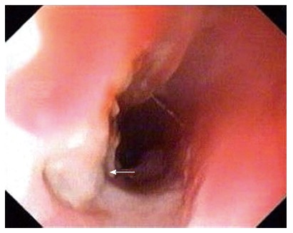
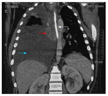
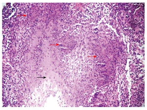
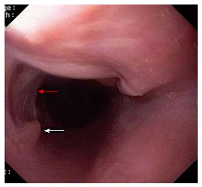
Esophageal tuberculosis is rare, constituting about 0.3% of gastrointestinal tuberculosis. It presents commonly with dysphagia, cough, chest pain in addition to fever and weight loss. Complications may include hemorrhage from the lesion, development of arterioesophageal fistula, esophagocutaneous fistula or tracheoesophageal fistula. There are very few reports of esophageal tuberculosis presenting with hematemesis due to ulceration. We report a patient with hematemesis that was due to the erosion of tuberculous subcarinal lymph nodes into the esophagus. A 15-year-old boy presented with hemetemesis as his only complaint. Esophagogastroduodenoscopy (EGD) revealed an eccentric ulcerative lesion involving 50% of circumference of the esophagus. Biopsy showed caseating epitheloid granulomas with lymphocytic infiltrates suggestive of tuberculosis. Computerised tomography of the thorax revealed thickening of the mid-esophagus with enlarged mediastinal lymph nodes in the subcarinal region compressing the esophagus along with moderate right sided pleural effusion. Patient was treated with anti-tuberculosis therapy (Rifampicin, Isoniazid, Pyrazinamide, Ethambutol) for 6 mo. Repeat EGD showed scarring and mucosal tags with complete resolution of the esophageal ulcer.
Keywords: Esophageal tuberculosis; Esophagogastroduodenoscopy; Hematemesis.
Figures
References
-
- Marshall JB. Tuberculosis of the gastrointestinal tract and peritoneum. Am J Gastroenterol. 1993;88:989–999. - PubMed
-
- Upadhyay AP, Bhatia RS, Anbarasu A, Sawant P, Rathi P, Nanivadekar SA. Esophageal tuberculosis with intramural pseudodiverticulosis. J Clin Gastroenterol. 1996;22:38–40. - PubMed
-
- Newman RM, Fleshner PR, Lajam FE, Kim U. Esophageal tuberculosis: a rare presentation with hematemesis. Am J Gastroenterol. 1991;86:751–755. - PubMed
-
- Fang HY, Lin TS, Cheng CY, Talbot AR. Esophageal tuberculosis: a rare presentation with massive hematemesis. Ann Thorac Surg. 1999;68:2344–2346. - PubMed
LinkOut - more resources
Full Text Sources
Other Literature Sources
Molecular Biology Databases
