Posterior mediastinal tuberculous lymphadenitis with dysphagia as the main symptom: a case report and literature review
- PMID: 24255790
- PMCID: PMC3815737
- DOI: 10.3978/j.issn.2072-1439.2013.09.03
Posterior mediastinal tuberculous lymphadenitis with dysphagia as the main symptom: a case report and literature review
Abstract
Introduction: Mediastinal tuberculous lymphadenitis (MTL) is mostly seen in primary tuberculosis in children, uncommon observed in adults. It usually presents the toxic symptoms of tuberculosis but rarely with symptoms characteristic of esophageal compression, such as dysphagia. Such patients can easily be misdiagnosed as esophageal neoplasm and get delayed or faulty treatment.
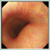
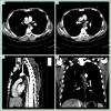
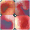
Case report: A 32-year-old man presented with dull chest pain of one month and dysphagia of five days. CRP was elevated, and a skin test was strongly positive. At upper endoscopy, a protruding lesion covered by normal mucosa was seen at 26 cm from the upper incisor. Barium swallow showed visible external compressive stricture on the middle-lower esophagus with normal mucosal pattern. Chest computed tomography (CT) scan showed a subcarinal mass adjacent to the esophageal wall in posterior mediastinum. An endoscopic ultrasonography (EUS) revealed a hypoechoic lesion suspected of esophageal stromal tumor in the fourth layer. A tissue was obtained by ultrasound-guided fine-needle aspiration (EUS-FNA), but cytopathology, bacilliculture and PCR test had no special findings. The patient required experimental antitubercular treatment and the protruding lesion shrank gradually during therapy period.
Conclusions: MTL could not be ignored in the differential diagnosis of posterior mediastinal mass with dysphagia. Analyzing and evaluating test results comprehensively is the key to make correct diagnosis and timely treatment. The experimental antituberculous treatment should be used if MTL is highly suspected.
Keywords: Mediastinal; dysphagia; tuberculous lymphadenitis.
Figures


Similar articles
-
Mediastinal tuberculous lymphadenitis presenting as a mediastinal mass with Dysphagia: a case report.Iran J Radiol. 2011 Sep;8(2):107-11. Epub 2011 Sep 25. Iran J Radiol. 2011. PMID: 23329926 Free PMC article.
-
Dysphagia due to mediastinal tuberculous lymphadenitis presenting as an esophageal submucosal tumor: a case report.Yonsei Med J. 1995 Sep;36(4):386-91. doi: 10.3349/ymj.1995.36.4.386. Yonsei Med J. 1995. PMID: 7483683
-
The Diagnosis of Esophageal Tuberculosis through an Endoscopic Ultrasound-guided Fine-needle Aspiration Biopsy.Intern Med. 2024 Sep 1;63(17):2399-2405. doi: 10.2169/internalmedicine.2824-23. Epub 2024 Feb 5. Intern Med. 2024. PMID: 38311428 Free PMC article.
-
Mediastinal tuberculous lymphadenitis presenting with insidious back pain in a male adult: a case report and review of the literature.J Int Med Res. 2021 Jan;49(1):300060520987102. doi: 10.1177/0300060520987102. J Int Med Res. 2021. PMID: 33445984 Free PMC article. Review.
-
The complete ''medical'' mediastinoscopy (EUS-FNA + EBUS-TBNA).Minerva Med. 2007 Aug;98(4):331-8. Minerva Med. 2007. PMID: 17921946 Review.
Cited by
-
An unusual cause of posterior mediastinal cyst.Lung India. 2015 Nov-Dec;32(6):609-10. doi: 10.4103/0970-2113.168123. Lung India. 2015. PMID: 26664169 Free PMC article.
-
Clinical Course of Patients With Mediastinal Lymph Node Tuberculosis and Risk Factors for Paradoxical Responses.J Korean Med Sci. 2023 Dec 4;38(47):e348. doi: 10.3346/jkms.2023.38.e348. J Korean Med Sci. 2023. PMID: 38050909 Free PMC article.
-
Is SUVmax Helpful in the Differential Diagnosis of Enlarged Mediastinal Lymph Nodes? A Pilot Study.Contrast Media Mol Imaging. 2018 Oct 28;2018:3417190. doi: 10.1155/2018/3417190. eCollection 2018. Contrast Media Mol Imaging. 2018. PMID: 30510493 Free PMC article.
-
Mediastinal lymphadenopathy: Causes, symptoms and factors predicting good yield of endoscopic ultrasound-guided biopsy.World J Clin Cases. 2025 Aug 6;13(22):105596. doi: 10.12998/wjcc.v13.i22.105596. World J Clin Cases. 2025. PMID: 40771743 Free PMC article.
References
-
- Venkateswaran RV, Barron DJ, Brawn WJ, et al. A forgotten old disease: mediastinal tuberculous lymphadenitis in children. Eur J Cardiothorac Surg 2005;27:401-4 - PubMed
-
- Amorosa JK, Smith PR, Cohen JR, et al. Tuberculous mediastinal lymphadenitis in the adult. Radiology 1978;126:365-8 - PubMed
-
- Argüello L.Endoscopic ultrasonography in submucosal lesions and extrinsic compressions of the gastrointestinal tract. Minerva Med 2007;98:389-93 - PubMed
-
- Motoo Y, Okai T, Ohta H, et al. Endoscopic ultrasonography in the diagnosis of extraluminal compressions mimicking gastric submucosal tumors. Endoscopy 1994;26:239-42 - PubMed
-
- Rösch T.Endoscopic ultrasonography in upper gastrointestinal submucosal tumors: a literature review. Gastrointest Endosc Clin N Am 1995;5:609-14 - PubMed
Publication types
LinkOut - more resources
Full Text Sources
Research Materials
Miscellaneous
