. 2014 Jan 9;123(2):208-16.
doi: 10.1182/blood-2013-03-489732. Epub 2013 Nov 19.

CD169 mediates the capture of exosomes in spleen and lymph node

Affiliations

CD169 mediates the capture of exosomes in spleen and lymph node

Sarah C Saunderson et al. Blood. .

Abstract

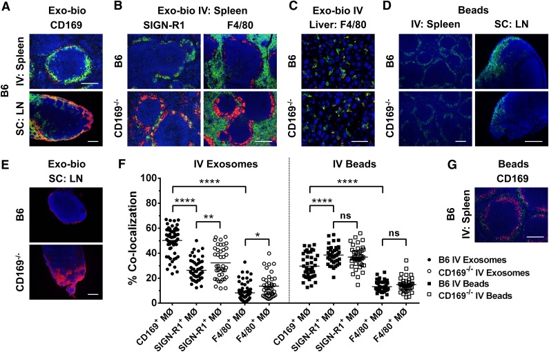
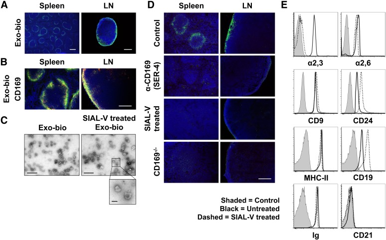
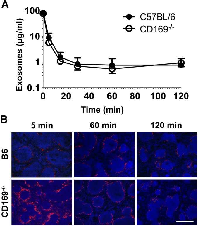
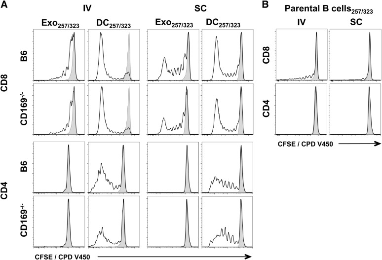
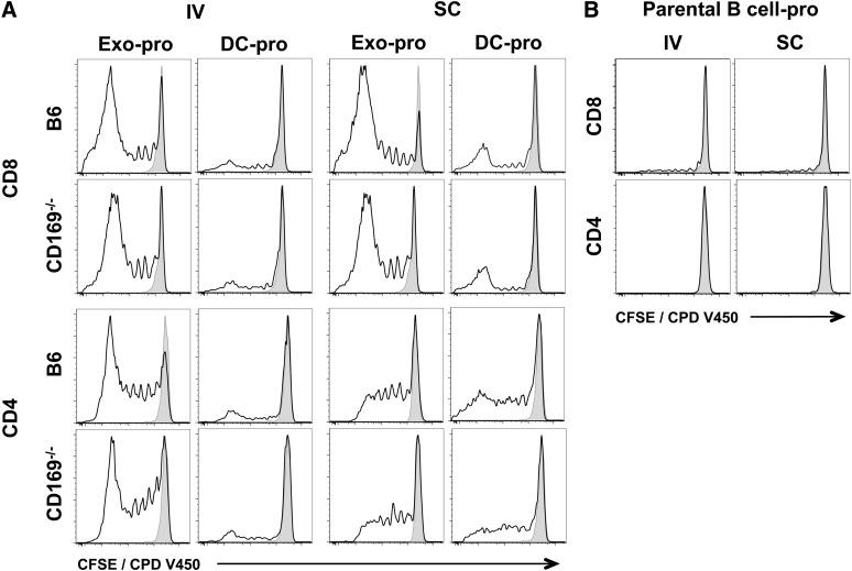
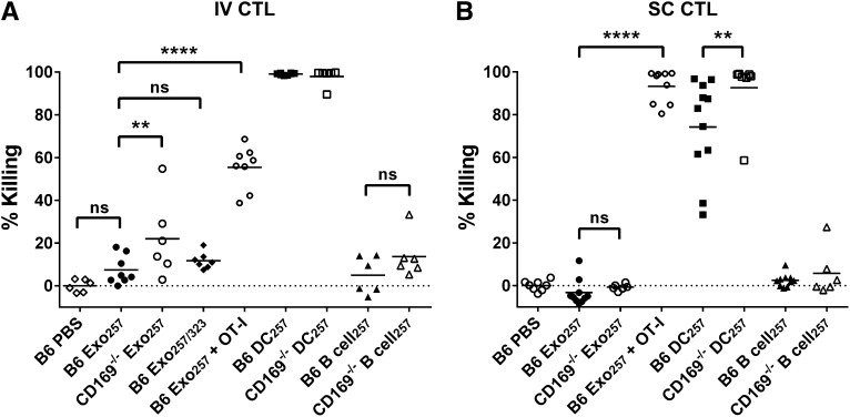
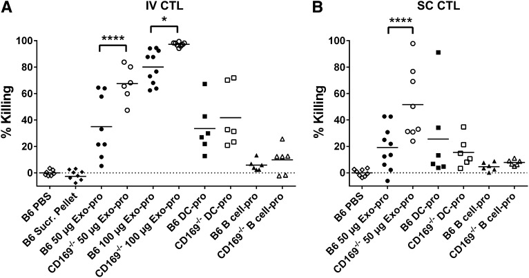
Exosomes are lipid nanovesicles released following fusion of the endosoma limiting membrane with the plasma membrane; however, their fate in lymphoid organs after their release remains controversial. We determined that sialoadhesin (CD169; Siglec-1) is required for the capture of B cell-derived exosomes via their surface-expressed α2,3-linked sialic acids. Exosome-capturing macrophages were present in the marginal zone of the spleen and in the subcapsular sinus of the lymph node. In vitro assays performed on spleen and lymph node sections confirmed that exosome binding to CD169 was not solely due to preferential fluid flow to these areas. Although the circulation half-life of exosomes in blood of wild-type and CD169(-/-) mice was similar, exosomes displayed altered distribution in CD169(-/-) mice, with exosomes freely accessing the outer marginal zone rim of SIGN-R1(+) macrophages and F4/80(+) red pulp macrophages. In the lymph node, exosomes were not retained in the subcapsular sinus of CD169(-/-) mice but penetrated deeper into the paracortex. Interestingly, CD169(-/-) mice demonstrated an enhanced response to antigen-pulsed exosomes. This is the first report of a role for CD169 in the capture of exosomes and its potential to mediate the immune response to exosomal antigen.


Figures

Figure 1
Figure 1
Aberrant distribution of exosomes in lymphoid tissues of CD169−/− in vivo. C57BL/6 or CD169−/− mice were (A [spleen],B,C,F) intravenously (IV) or (A [LN],E) subcutaneously (SC) injected in the forelimb with 100 or 50 µg of biotinylated B cell–derived exosomes (Exo-bio; purified by ultracentrifugation), respectively. (D,F,G) Alternatively, C57BL/6 or CD169−/− mice were IV or SC injected with 2 × 10¹¹ or 1 × 10¹¹ 100 nm fluorescent microspheres, respectively (green). For IV or SC routes, mice were killed at 5 minutes with spleens and livers harvested or at 30 min and draining LN harvested, respectively. (A-C,E) Exo-bio was detected with streptavidin-Alexa-594 (red). Sections were colabeled for (A,G) marginal metallophilic or subcapsular sinus macrophages with anti-CD169 (MOMA-1), (B) MZ or red pulp macrophages with anti–SIGN-R1 (ER-TR9) or anti-F4/80, respectively, or (C) Kupffer cells with anti-F4/80. Primary antibodies were detected with (A-C) anti-rat IgG-Alexa-488 (green) or (G) anti-rat IgG-Alexa-594 (red), and nuclei were counterstained with DAPI (blue). Original magnification, (A [LN],D[spleen],E) ×100 , (A [spleen],B,D [LN],G) ×200, and (C) ×400. Bar represents (A [spleen],B,D [LN],G) 200 µm, (A [LN],D [spleen],E) 250 µm, and (C) 50 µm. All results are representative of ≤4 mice per group. (F) Percent colocalization was calculated from fluorescent microscopy photos of (A [spleen],B,G) spleen sections. Ten individual photographs per mouse (original magnification, ×200) were analyzed for colocalization of green (Alexa-488) and red (Alexa-594) signal using the Manders’ coefficient with ImageJ. Each point represents an individual photograph; line indicates mean. Circles, exosomes; squares, beads; closed symbols, C57BL/6 mice; open symbols, CD169−/− mice. One-way ANOVA with Bonferroni postcorrection test was performed: ns, not significant; *P < .05; **P < .01; ****P < .0001.
Figure 2
Figure 2
Exosomes are bound by CD169+ macrophages in the spleen and LN in the absence of blood or lymph flow. Exo-bio was applied to naïve C57BL/6 or CD169−/− spleen and LN sections using a modified Stamper-Woodruff assay. Biotin was detected with (A) streptavidin-Alexa-488 (green) or (B) -Alexa-594 (red) and marginal metallophilic or subcapsular sinus macrophages stained with anti-CD169 (MOMA-1). Primary antibody was detected with (B) anti-rat IgG-Alexa-488 (green) and nuclei counterstained with DAPI (blue). (C) Exo-bio ± treatment with Vibrio cholerae–derived sialidase (α2,3-linked sialic acid preferential cleavage [SIAL-V]) were purified using a sucrose cushion, negatively stained and visualized by transmission electron microscopy. No apparent differences in morphology were observed between samples; diameter range was 70 to 120 nm. Photographs are representative of the preparations as a whole. (D) Exo-bio was bound to naïve C57BL/6 sections in the presence of negative control antibody or CD169 neutralizing antibody (SER-4). Alternatively, Exo-bio or SIAL-V–treated Exo-bio was applied to naïve C57BL/6 or CD169−/− sections, respectively. Exosomes and nuclei were detected as described in A. (E) Exosomes ± SIAL-V treatment was bound to aldehyde-sulfate microspheres and analyzed by flow cytometry for α2,3- and α2,6-linked sialic acid expression using biotinylated MAL-II and SNA lectins, respectively. In addition, CD9, CD24, MHC-II, CD19, immunoglobulin, and CD21 expression was measured. Shaded peak, negative control (BSA-conjugated aldehyde-sulfate microspheres); black line, untreated exosomes; dashed line, SIAL-V–treated exosomes. Results representative of ≥3 separate experiments and/or exosome preparations, with (D:LN) LN sections from ≥4 anatomically distinct locations per experiment. Original magnification, (A [spleen]) ×40, (A [LN]) ×100, (B,D) ×200, (C [Exo-bio]) ×24 500, and (C [SIAL-V–treated Exo-bio]) ×17 500. Bar represents (A [spleen]) 500 µm, (A [LN]) 250 µm, (B,D) 200 µm, (C) 500 nm, and (C, inset) 100 nm.
Figure 3
Figure 3
Exosome clearance and distribution in vivo. (A) C57BL/6 or CD169−/− mice were anesthetized and then IV injected with 100 µg Exo-bio (purified by ultracentrifugation). Mice were tail bled at the indicated time points. MHC-II+ exosome concentration was analyzed by enzyme-linked immunosorbent assay from plasma samples; Exo-bio spiked plasma was used as a standard. Closed circles, C57BL/6 mice; open circles, CD169−/− mice. (B) C57BL/6 or CD169−/− mice were IV injected with 100 µg Exo-bio. Mice were killed at the indicated time points, and spleens were harvested. Exo-bio was detected with streptavidin-Alexa-594 and nuclei counterstained with DAPI. (B) Original magnification, ×100. Bar represents 500 µm. Results representative of (A,B) 6 mice (120 minutes) and (B) 3 to 6 mice (5 and 60 minutes).
Figure 4
Figure 4
In vivo T-cell proliferation in response to exosomal-peptide antigen. C57BL/6 or CD169−/− mice were immunized IV or SC in the forelimb with (A) PBS, 100 µg sucrose cushion purified Exo257/323, and 105 DC257/323 or (B) 105 parental B cell257/323. Exosomes and cells were all pulsed simultaneously with 1 μM ovalbumin peptides OVA257-264 and OVA323-339. T-cell proliferation of adoptively cotransferred OT-I (CD8) and OT-II (CD4) cells (CFSE or CPD V450) were analyzed 5 days after immunization by flow cytometry. Black line, test group; shaded peak, PBS-immunized mice. Results representative of ≥6 mice per group.
Figure 5
Figure 5
T-cell proliferation in response to exosomal-protein antigen. C57BL/6 or CD169−/− mice were immunized IV or SC in the forelimb with (A) PBS, 50 µg sucrose cushion purified exosomes derived from B cells cultured with 200 µg/mL ovalbumin protein for 2 days (Exo-pro), and 105 DC-pro or (B) 105 parental B cell-pro. DC and B cells were cultured with 200 µg/mL ovalbumin protein for 2 days. T-cell proliferation of adoptively cotransferred OT-I (CD8) and OT-II (CD4) cells (CFSE or CPD V450) were analyzed 5 days after immunization by flow cytometry. Black line, test group; shaded peak, PBS-immunized mice. Results representative of ≥6 mice per group.
Figure 6
Figure 6
Enhanced cytotoxic responses to intravenous exosomal-peptide in CD169−/− mice. C57BL/6 or CD169−/− mice were immunized (A) IV or (B) SC with PBS and 100 or 50 µg Exo257, respectively, 100 μg Exo257/323 (IV), 105 DC257, or 105 parental B cell257. Where stated, mice were supplemented IV with 107 OT-I splenocytes prior to immunization. Seven days after immunization, mice were adoptively transferred with unpulsed (CFSE low) or OVA257-264-pulsed (CFSE high) target cells. In vivo killing was analyzed 18 hours later by flow cytometry. Results representative of ≥6 mice per group using exosomes purified by ultracentrifugation. Line, mean. One-way ANOVA with Bonferroni postcorrection was performed: ns, not significant; **P < .01; ****P < .0001.
Figure 7
Figure 7
Enhanced cytotoxic responses to exosomal-protein in CD169−/− mice. C57BL/6 or CD169−/− mice were immunized (A) IV or (B) SC with PBS, pellet from exosome sucrose cushion purification (IV: B6 Sucr. Pellet), 50 μg sucrose cushion purified Exo-pro or 100 µg Exo-pro (purified by ultracentrifugation), 105 DC-pro, or 105 parental B cell-pro. Seven days after immunization, mice were adoptively transferred with unpulsed (CFSE low) or OVA257-264-pulsed (CFSE high) target cells. In vivo killing was analyzed 18 hours later by flow cytometry. Results representative of ≥6 mice per group. One-way ANOVA with Bonferroni postcorrection was performed: *P < .05; ****P < .0001.

Similar articles

Cited by

References

    1. Zitvogel L, Regnault A, Lozier A, et al. Eradication of established murine tumors using a novel cell-free vaccine: dendritic cell-derived exosomes. Nat Med. 1998;4(5):594–600. - PubMed
    1. André F, Chaput N, Schartz NE, et al. Exosomes as potent cell-free peptide-based vaccine. I. Dendritic cell-derived exosomes transfer functional MHC class I/peptide complexes to dendritic cells. J Immunol. 2004;172(4):2126–2136. - PubMed
    1. Nolte-’t Hoen EN, Buschow SI, Anderton SM, Stoorvogel W, Wauben MH. Activated T cells recruit exosomes secreted by dendritic cells via LFA-1. Blood. 2009;113(9):1977–1981. - PubMed
    1. Rieu S, Geminard C, Rabesandratana H, Sainte-Marie J, Vidal M. Exosomes released during reticulocyte maturation bind to fibronectin via integrin alpha4beta1. Eur J Biochem. 2000;267(2):583-590. - PubMed
    1. Wubbolts R, Leckie RS, Veenhuizen PT, et al. Proteomic and biochemical analyses of human B cell-derived exosomes. Potential implications for their function and multivesicular body formation. J Biol Chem. 2003;278(13):10963–10972. - PubMed

Publication types

MeSH terms