. 2014 Feb 21;9(2):459-67.
doi: 10.1021/cb4006744. Epub 2013 Nov 20.

Moenomycin resistance mutations in Staphylococcus aureus reduce peptidoglycan chain length and cause aberrant cell division

Affiliations

Moenomycin resistance mutations in Staphylococcus aureus reduce peptidoglycan chain length and cause aberrant cell division

Yuriy Rebets et al. ACS Chem Biol. .

Abstract

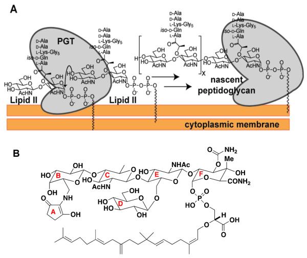
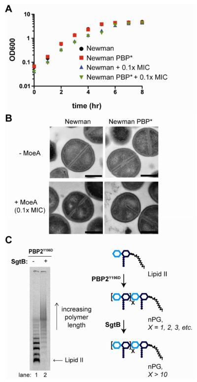
Staphylococcus aureus is a Gram-positive pathogen with an unusual mode of cell division in that it divides in orthogonal rather than parallel planes. Through selection using moenomycin, an antibiotic proposed to target peptidoglycan glycosyltransferases (PGTs), we have generated resistant mutants containing a single point mutation in the active site of the PGT domain of an essential peptidoglycan (PG) biosynthetic enzyme, PBP2. Using cell free polymerization assays, we show that this mutation alters PGT activity so that much shorter PG chains are made. The same mutation in another S. aureus PGT, SgtB, has a similar effect on glycan chain length. Moenomycin-resistant S. aureus strains containing mutated PGTs that make only short glycan polymers display major cell division defects, implicating PG chain length in determining bacterial cell morphology and division site placement.


Figures

Figure 1
Figure 1
S. aureus peptidoglycan glycosyltransferases (PGTs) catalyze elongation of the glycan chain. a) Schematic of the PGT reaction to produce nascent (uncrosslinked) peptidoglycan (nPG) from the precursor Lipid II. There are three PGTs in S. aureus: SgtA, SgtB, and PBP2. b) Structure of the antibiotic moenomycin A (MoeA), a PGT inhibitor, with ring nomenclature indicated.
Figure 2
Figure 2
MoeA resistance mutations are located in the catalytic cleft of the PGT domain of S. aureus PBP2. a) Surface representation of the PGT domain of PBP2 with bound MoeA (19). The catalytic glutamate, Glu114, defines the center of the active site and is shown in red. Tyr196 is shown in cyan and Pro234 in green. PDB ID: 2OLV. Image generated by PyMOL. b) Gel electrophoresis showing the length distributions of PG polymers produced from Lipid II in vitro by wild type PBP2 and the PBP2Y196D mutant enzymes as a function of time. c) Model illustrating how different product distributions can arise in wild type and mutant enzymes. PBP2 polymerizes Lipid II in a processive manner, while PBP2Y196D polymerizes by a distributive mechanism. In the key, GlcNAc is N-acetylglucosamine and MurNAc is N-acetylmuramic acid.
Figure 3
Figure 3
Moenomycin affects SgtB activity in the PBP2Y196D mutants. a) Growth of wild type S. aureus Newman and PBP2Y196D mutant (PBP*) cells in the absence and presence of 0.1X MIC MmA. Concentration of MmA is based on the MIC of each strain shown in Table S4. b) Representative transmission electron microscopy (TEM) images of S. aureus Newman and PBP* cells treated with 0.1X MIC of MoeA. The half septum defect is highlighted in the PBP* strain with added MoeA. Quantification of division defects for each strain is shown in Figure S5A. Scale bars indicate 500 nm. c) Gel electrophoresis shows that short PG polymers produced by PBP2Y196D are elongated by SgtB in vitro, as indicated by the scheme on the right. PBP2Y196D reactions (1 hr) were incubated in the absence or presence of StgB (30 min) prior to heat quenching.
Figure 4
Figure 4
Inability to produce long PG strands leads to aberrant cell division. a) Light microscopy quantification of cell division states during early exponential growth of the wild-type S. aureus Newman and RN4220 strains, of the mutant PBP* strains, and of the double mutant RN4220 PBP2*ΔsgtB strain. The percentage of cells with division aberrations is underestimated as only cells with clearly visible septa could be counted; cells buried in clumps, which may be the most affected, could not be counted. See Figure S6 for more details. b) Light microscopy of wild type S. aureus RN4220 and the double mutant RN4220 PBP2*ΔsgtB. The cell outline and septa are visualized with a membrane dye (TMA-DPH). The arrows indicate the groups described in (A): full septum (blue); forming septum (red), hemi-septum (purple); parallel septa (green); and otherwise aberrant septum (orange). Scale bar represents 2 μm. c) TEM of wild-type S. aureus RN4220 (left) and the double mutant RN4220 PBP2*ΔsgtB. The double mutants exhibit aberrant division phenotypes including chaining phenotype (middle) and hemi-septa formation in (right, indicated with arrow). Scale bar represents 1 μm. d) Model illustrating cell division of wild type versus mutant S. aureus cells. Dysregulated PG synthesis in the mutant cells leads to formation of chaining and hemi-septum phenotypes.

References

    1. Vollmer W, Blanot D, de Pedro MA. Peptidoglycan structure and architecture. FEMS Microbiol. Rev. 2008;32:149–167. - PubMed
    1. Sun SX, Jiang H. Physics of bacterial morphogenesis. Microbiol. Mol. Biol. Rev. 2011;75:543–565. - PMC - PubMed
    1. Typas A, Banzhaf M, Gross CA, Vollmer W. From the regulation of peptidoglycan synthesis to bacterial growth and morphology. Nat. Rev. Microbiol. 2012;10:123–136. - PMC - PubMed
    1. Sauvage E, Kerff F, Terrak M, Ayala JA, Charlier P. The penicillin-binding proteins: structure and role in peptidoglycan biosynthesis. FEMS Microbiol. Rev. 2008;32:234–258. - PubMed
    1. Lovering AL, Gretes M, Strynadka NCJ. Structural details of the glycosyltransferase step of peptidoglycan assembly. Curr. Opin. Struct. Biol. 2008;18:534–543. - PubMed

Publication types

MeSH terms