

Polyol effects on growth factors and MAPK signaling in rat retinal capillary cells
- PMID: 24256145
- PMCID: PMC4094127
- DOI: 10.1089/jop.2013.0170
Polyol effects on growth factors and MAPK signaling in rat retinal capillary cells
Abstract
Purpose: Recent studies report that growth factor and signaling changes in rat lenses do not directly result from the presence of diabetes or sorbitol/galactitol (polyol) formation/accumulation, but from secondary osmotic changes associated with the aldose reductase (AR) catalyzed polyol formation. AR is also present in rat retinal pericyte and endothelial cells; however, significant polyol formation only occurs in pericytes and this does not appear to be linked to osmotic changes. The purpose of this study was to determine whether polyol formation and AR activity are similarly linked to growth factor and signaling changes in the rat capillary cells despite the apparent absence of osmotic stress.
Methods: Conditionally immortalized rat retinal pericyte (TR-rPCT) and endothelial (TR-iBRB) cell lines were cultured on collagen type 1-coated dishes in the DMEM containing 5.5 mM glucose. After 24 h of initial culture, the medium was replaced with a serum-free medium containing 5.5, 25, or 50 mM glucose or galactose with/without the aldose reductase inhibitors (ARIs) AL1576 or tolrestat for periods of up to 48 h. Growth factors and transduction pathways were measured by Western blots using the antibodies against basic FGF, IGF-1, TGF-β, P-ERK1/2, P-SAPK/JNK, and P-Akt.
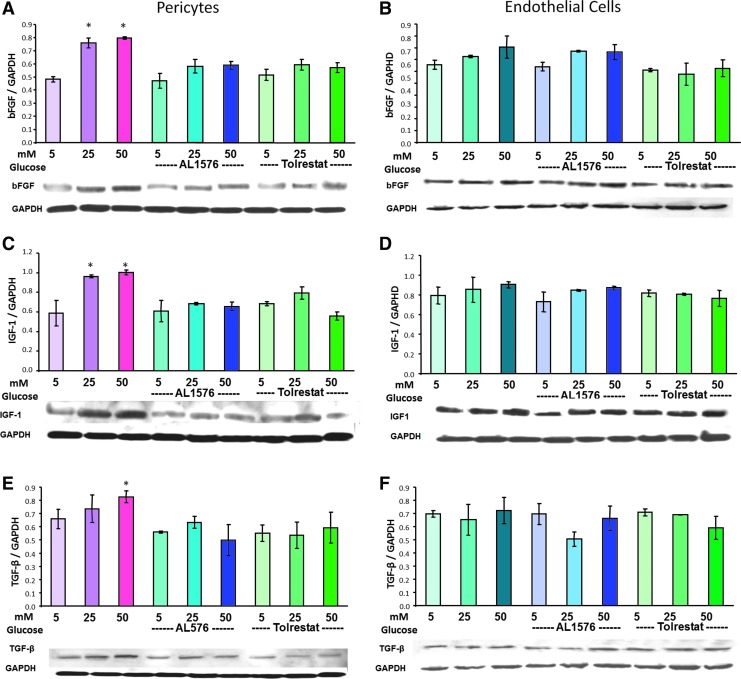
Results: Sorbitol accumulation was only observed in pericytes, while galactitol was present in both pericytes and endothelial cells. Pericytes cultured in high glucose showed increased expression of the growth factors basic FGF, IGF-1, TGF-β, and signaling in P-Akt, P-ERK1/2, and P-SAPK/JNK compared with those cultured in 5.5 mM glucose and these expressions were normalized by the presence of ARIs. Similar results were observed with galactose media. In contrast, endothelial cells cultured in high glucose media showed neither growth factor or signaling changes. In galactose media, endothelial cells showed increased expression of basic FGF, IGF-1, TGF-β, P-ERK1/2, and P-SAPK/JNK, which were only partially reduced by ARIs.
Conclusion: Growth factor and MAPK signaling expression in pericytes are linked to the presence of polyols. Pericytes, which readily accumulate sorbitol/galactitol that is inhibited by ARIs, show expression changes similar to those observed in rat lenses. In contrast, endothelial cells only show partial expression changes that are linked to galactitol accumulation.
Figures


Similar articles
-
Polyol formation in cell lines of rat retinal capillary pericytes and endothelial cells (TR-rPCT and TR-iBRB).J Ocul Pharmacol Ther. 2009 Aug;25(4):299-308. doi: 10.1089/jop.2008.0070. J Ocul Pharmacol Ther. 2009. PMID: 19450153 Free PMC article.
-
Osmotic stress, not aldose reductase activity, directly induces growth factors and MAPK signaling changes during sugar cataract formation.Exp Eye Res. 2012 Aug;101:36-43. doi: 10.1016/j.exer.2012.05.007. Epub 2012 Jun 15. Exp Eye Res. 2012. PMID: 22710095 Free PMC article.
-
Response of rat retinal capillary pericytes and endothelial cells to glucose.J Ocul Pharmacol Ther. 2011 Feb;27(1):7-15. doi: 10.1089/jop.2010.0051. Epub 2010 Nov 20. J Ocul Pharmacol Ther. 2011. PMID: 21091050 Free PMC article.
-
Aldose reductase in diabetic complications of the eye.Metabolism. 1979 Apr;28(4 Suppl 1):462-9. doi: 10.1016/0026-0495(79)90057-x. Metabolism. 1979. PMID: 45423 Review.
-
Aldose reductase / polyol inhibitors for diabetic retinopathy.Curr Pharm Biotechnol. 2011 Mar 1;12(3):373-85. doi: 10.2174/138920111794480642. Curr Pharm Biotechnol. 2011. PMID: 20939801 Review.
Cited by
-
Novel transgenic mouse models develop retinal changes associated with early diabetic retinopathy similar to those observed in rats with diabetes mellitus.Exp Eye Res. 2014 Feb;119:77-87. doi: 10.1016/j.exer.2013.12.009. Epub 2013 Dec 25. Exp Eye Res. 2014. PMID: 24370601 Free PMC article.
-
Effects of microRNA‑217 on high glucose‑induced inflammation and apoptosis of human retinal pigment epithelial cells (ARPE‑19) and its underlying mechanism.Mol Med Rep. 2019 Dec;20(6):5125-5133. doi: 10.3892/mmr.2019.10778. Epub 2019 Oct 30. Mol Med Rep. 2019. PMID: 31702814 Free PMC article.
-
Repression of retinal microvascular endothelial cells by transthyretin under simulated diabetic retinopathy conditions.Int J Ophthalmol. 2016 Jun 18;9(6):809-15. doi: 10.18240/ijo.2016.06.03. eCollection 2016. Int J Ophthalmol. 2016. PMID: 27366679 Free PMC article.
-
Antioxidant and Anti-Inflammatory Effects of Blueberry Anthocyanins on High Glucose-Induced Human Retinal Capillary Endothelial Cells.Oxid Med Cell Longev. 2018 Feb 22;2018:1862462. doi: 10.1155/2018/1862462. eCollection 2018. Oxid Med Cell Longev. 2018. PMID: 29682153 Free PMC article.
-
Transthyretin represses neovascularization in diabetic retinopathy.Mol Vis. 2016 Oct 12;22:1188-1197. eCollection 2016. Mol Vis. 2016. PMID: 27746673 Free PMC article.
References
-
- Progression of retinopathy with intensive versus conventional treatment in the Diabetes Control and Complications Trial. Diabetes Control and Complications Trial Research Group. Ophthalmology. 102:647–661, 1995 - PubMed
-
- The U.K. Prospective Diabetes Study Group. Intensive blood glucose control with sulphonylureas or insulin compared with conventional treatment and risk of complications in patients with type 2 diabetes. Lancet. 352:837–853, 1998 - PubMed
-
- Kalichman M.W., et al. . Nerve conduction velocity, laser Doppler flow, and axonal caliber in galactose and streptozotocin diabetes. Brain Res. 810:130–137, 1998 - PubMed
-
- Cogan D.G., et al. . NIH conference. Aldose reductase and complications of diabetes. Ann Intern Med. 101:82–91, 1984 - PubMed
-
- Robison W.G.Galactosemic animal models. In: Sima A.A.F., and Shafir E., eds. Aniaml Models in Diabetes A Primer, series: Frontiers in Aminal Diabetes Research. Amsterdam: Harwood Academic Publishers; 2000; pp. 273–308
Publication types
MeSH terms
Substances
Grants and funding
LinkOut - more resources
Full Text Sources
Other Literature Sources
Research Materials
Miscellaneous
