. 2014 Feb;281(3):696-707.
doi: 10.1111/febs.12615. Epub 2013 Dec 12.

Insights into the suppressor of T-cell receptor (TCR) signaling-1 (Sts-1)-mediated regulation of TCR signaling through the use of novel substrate-trapping Sts-1 phosphatase variants

Affiliations

Insights into the suppressor of T-cell receptor (TCR) signaling-1 (Sts-1)-mediated regulation of TCR signaling through the use of novel substrate-trapping Sts-1 phosphatase variants

Boris S Luis et al. FEBS J. 2014 Feb.

Abstract

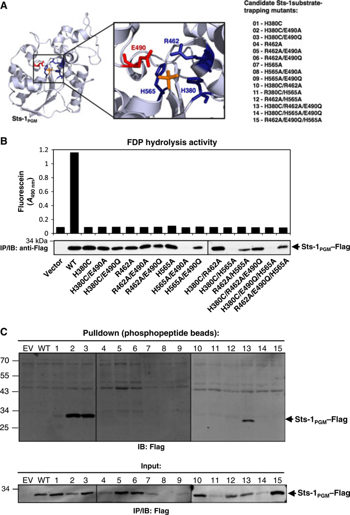
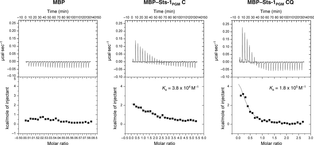
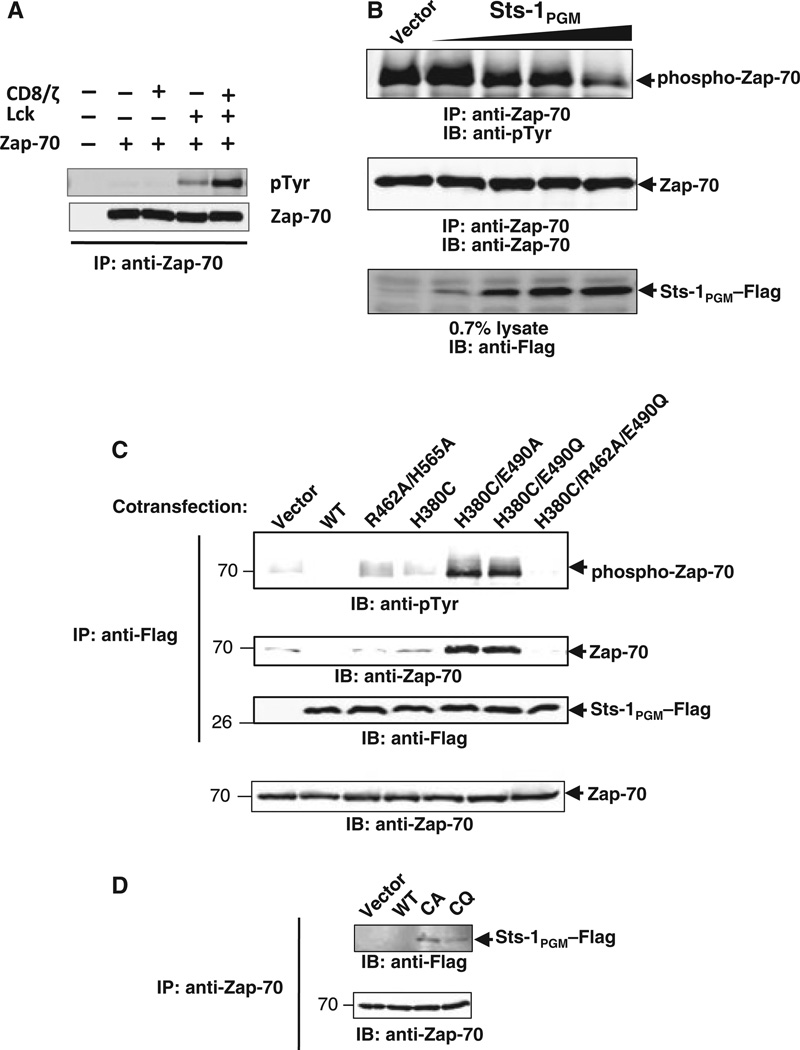
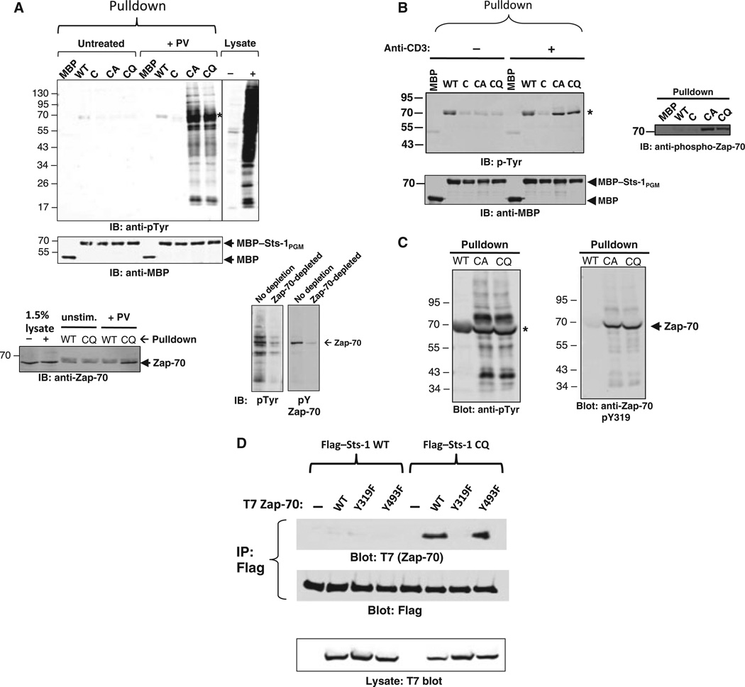
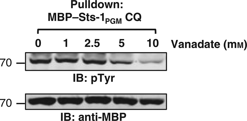
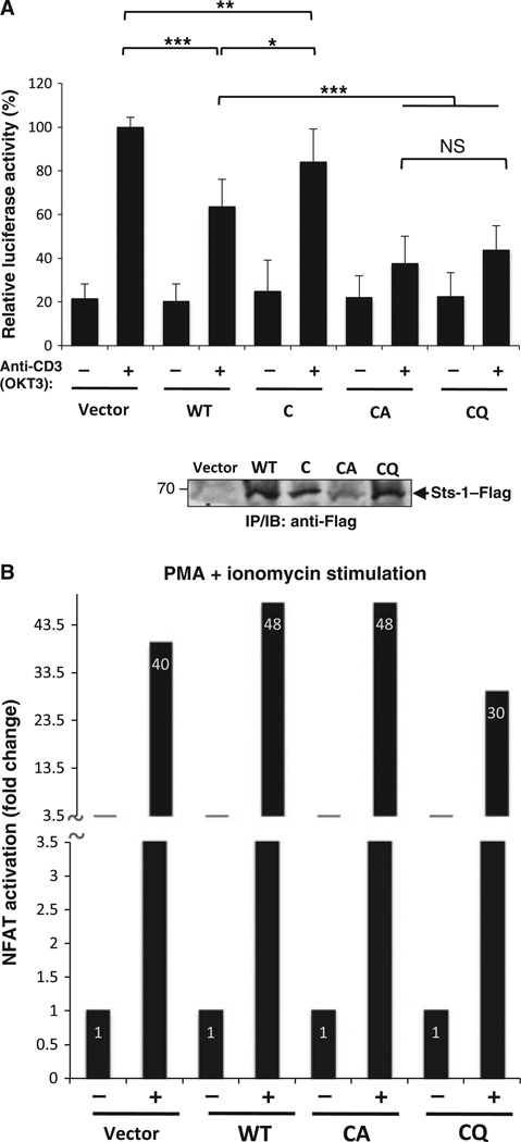
High affinity substrate-trapping protein tyrosine phosphatases have been widely used both to investigate the endogenous targets of many phosphatases and to address questions of substrate specificity. Herein, we extend the concept of a substrate-trapping phosphatase to include an enzyme of the histidine phosphatase superfamily. This is the first description of substrate-trapping technology applied to a member of the histidine phosphatase family. The phosphatase suppressor of T-cell receptor signaling (Sts)-1 has recently been reported to negatively regulate signaling downstream of the T-cell receptor. We generated high-affinity substrate-trapping variants of Sts-1 by mutagenesis of key active site residues within the phosphatase catalytic domain. Mutation of both the nucleophilic His380 and the general acid Glu490 yielded Sts-1 enzymes that were catalytically inactive but showed high affinity for an important tyrosine kinase in T cells that Sts-1 is known to regulate, Zap-70. Sts-1 substrate-trapping mutants isolated tyrosine-phosphorylated Zap-70 from lysates of activated T cells, validating Zap-70 as a possible substrate for Sts-1 and highlighting the efficacy of the mutants as substrate-trapping agents. Inhibition of the Zap-70 interaction by vanadate suggests that the substrate-trapping effect occurred via the Sts-1 phosphatase active site. Finally, overexpression of Sts-1 substrate-trapping mutants in T cells blocked T-cell receptor signaling, confirming the inhibitory effect of Sts-1 on Zap-70.

Keywords: T-cell receptor (TCR) signaling; histidine phosphatase superfamily; suppressor of T-cell receptor signaling (Sts) proteins.


Conflict of interest statement

The authors declare no financial conflict of interest.

Figures

Fig. 1
Fig. 1
Development of Sts-1 high-affinity mutants. (A) Representation of conserved active site residues in Sts-1PGM, generated in PYMOL with the crystal structure of Sts-1PGM complexed with phosphate (Protein Data Bank ID: 2IKQ). A total of 15 candidate Sts-1 substrate-trapping mutants were generated by site-directed mutagenesis of His380 to cysteine (H380C), Arg462 to alanine (R462A), and His565 to alanine (H565A), singly or in combination with mutations targeting the proton donor Glu490 (E490A or E490Q). (B) Phosphatase activity assay of candidate substrate-trapping mutants. Flag-tagged wild-type (WT) or mutant Sts-1PGM was expressed in HEK293T cells, immunoprecipitated with antibody against Flag, immobilized on protein A–Sepharose beads, and evaluated for FDP phosphatase activity. The FDP assay was carried out at 37 °C for 15 min with 0.5 mm FDP, and this was followed by detection of the fluorescein product in the supernatants by measuring the absorbance at 490 nm. Sts-1PGM expression levels were evaluated by immunoblotting (IB) with mAb against Flag. (C) A phosphopeptide pulldown assay with lysates of HEK293T cells transfected with empty vector (EV), wild-type Sts-1PGM (WT), or mutant Sts-1PGMs corresponding to the list in (A) (lanes 1–15). Protein complexes were resolved by SDS/PAGE and immunoblotted with antibodies against Flag. The level of Sts-1PGM in the input was determined by analysis of a fraction of the lysate used in the pulldown assay (lower blot). As controls, empty beads (agarose blocked with ethanolamine) were used against an equal volume of the HEK293T lysates; they showed no interaction (not shown). IP, immunoprecipitation.
Fig. 2
Fig. 2
Sts-1PGM CQ stably interacts with a small-molecule substrate. Calorimetric titrations of purified recombinant MBP (left), MBP–Sts-1PGM C (middle) and MBP–Sts-1PGM CQ (right) with OMFP. The binding affinities obtained from this analysis are indicated: Sts-1PGM C, Ka = 3.8 × 10⁴ m−1 ; Sts-1PGM CQ, Ka = 1.75 × 10⁵ m−1.
Fig. 3
Fig. 3
Interaction of Sts-1PGM high-affinity mutants and the T-cell tyrosine kinase Zap-70. (A) HEK293T cells were transfected with cDNA encoding Zap-70, singly or in combination with constructs encoding Lck and CD8/ζ (a chimeric protein containing the extracellular and transmembrane domains of the CD8 coreceptor and the cytoplasmic domain of TCRζ). Anti-Zap-70 immunoprecipitates were immunoblotted against mAb against pTyr (4G10) to determine phosphorylation levels, and against antibody against Zap-70 to verify equal Zap-70 expression in cells. (B) Expression plasmids for Zap-70, CD8/ζ and Lck were cotransfected into HEK293T cells along with either empty vector or constructs encoding Sts-1PGM at increasing plasmid DNA concentrations. Anti-Zap-70 immunoprecipitates were analyzed by immunoblotting to determine Zap-70 tyrosine dephosphorylation by Sts-1PGM. (C) HEK293T cells were transfected with Zap-70, Lck, and CD8/ζ, along with Flag-tagged wild-type Sts-1PGM or mutants. Twenty-four hours post-transfection, cells were lysed, and protein complexes were assessed by immunoprecipitation (IP)/immunoblot (IB) analysis. Flag immunoprecipitates were immunoblotted with antibodies against pTyr and Zap-70 to detect Zap-70, and with antibodies against Flag to assess Sts-1PGM levels. Levels of Zap-70 were assessed by IP/IB analysis (lower panel). (D) The converse IP analysis detected Sts-1PGM coprecipitating with antibodies against Zap-70.
Fig. 4
Fig. 4
Isolation of tyrosine-phosphorylated proteins by high-affinity Sts-1PGM mutants. (A) Lysates of Jurkat T cells treated with pervanadate (+PV) or left untreated were incubated with equal levels of MBP or MBP fusions of wild-type Sts-1PGM (WT), Sts-1PGM C, and the high-affinity mutants Sts-1PGM CA and Sts-1PGM CQ that had been immobilized on amylose beads. Bound-tyrosine phosphorylated proteins were analyzed by anti-pTyr immunoblotting (IB). The last two lanes contain 12.5% of untreated (−) and treated (+) Jurkat lysates. The prominent 70-kDa tyrosine-phosphorylated band that binds to Sts-1PGM CA/Sts-1PGM CQ contains Zap-70, as revealed by IB analysis with specific antibodies (bottom left), and depletion analysis in which Zap-70 was depleted from the lysate prior to Sts-1PGM CQ pulldown (bottom right). (B) Similar analysis as in (A), performed with lysates derived from TCR-stimulated Jurkat T cells. Phosphorylated Zap-70 was recognized with a cocktail of phosphorylation site-specific antibodies recognizing Zap-70 pTyr319, pTyr292, and pTyr493. (C) Similar analysis as in (A), performed with lysates derived from TCR-stimulated primary mouse T cells isolated from Sts-1/2−/− mice. (D) Interaction of Sts-1PGM CQ is abrogated in the absence of Zap-70 Tyr319 phosphorylation. Full-length wild-type Sts-1 and Sts-1PGM CQ were coexpressed with the indicated Zap-70 mutants, and evaluated for trapping abilities. The data presented are representative of three independent experiments. IP, immunoprecipitation.
Fig. 5
Fig. 5
Vanadate inhibits the stable interaction of Sts-1PGM CQ with phosphorylated Zap-70. Complex formation between the high-affinity Sts-1PGM mutant and phosphorylated Zap-70 in the presence of increasing concentrations of vanadate was assessed by pulldown assay. Samples were analyzed by immunoblotting (IB) with antibodies against pTyr and MBP.
Fig. 6
Fig. 6
High-affinity substrate-trapping mutants inhibit TCR signaling. (A) Jurkat T cells were cotransfected with an NFAT-luciferase reporter construct, and plasmids encoding either Flag-tagged, full-length wild-type Sts-1 (WT), Sts-1PGM C, Sts-1PGM CA, or Sts-1PGM CQ. Following 8 h of TCR stimulation, cells were harvested, and reporter activity was determined. Error bars are standard deviations from three to four independent experiments. *P < 0.05, **P < 0.01, and ***P < 0.005, where P is the probability associated with a Student's paired t-test, with a two-tailed distribution. NS, not significant. (B) High-affinity Sts-1 mutants fail to regulate signaling when the TCR is bypassed. Jurkat cells were transfected with a firefly luciferase reporter construct under the control of NFAT binding sequences, a Renilla luciferase reporter construct under the control of a constitutive promoter, and plasmids encoding Flag-tagged, full-length wild-type Sts-1 (WT), or the substrate-trapping mutants Sts-1PGM CA or Sts-1PGM CQ. At least 20 h post-transfection, the cells were stimulated with PMA (50 ng mL−1) plus 1 µm ionomycin. Levels of NFAT reporter activation were analyzed by luciferase assay. Bar graphs represent luciferase activities relative to unstimulated Jurkat transfections.

Similar articles

Cited by

References

    1. Smith-Garvin JE, Koretzky GA, Jordan MS. T cell activation. Annu Rev Immunol. 2009;27:591–619. - PMC - PubMed
    1. Mustelin T, Tasken K. Positive and negative regulation of T-cell activation through kinases and phosphatases. Biochem J. 2003;371:15–27. - PMC - PubMed
    1. Samelson LE. Immunoreceptor signaling. In: Cantley L, Hunter T, Sever R, Thorner J, editors. Cold Spring Harbor Perspectives in Biology: Signal Transduction. Woodbury, NY: Cold Spring Harbor Laboratory Press; 2011.
    1. Love PE, Hayes SM. ITAM-mediated signaling by the T-cell antigen receptor. In: Samelson LE, Shaw A, editors. Cold Spring Harbor Perspectives in Biology: Immunoreceptor Signaling. Woodbury, NY: Cold Spring Harbor Laboratory Press; 2010. - PMC - PubMed
    1. Deindl S, Kadlecek TA, Brdicka T, Cao X, Weiss A, Kuriyan J. Structural basis for the inhibition of tyrosine kinase activity of Zap-70. Cell. 2007;129:735–746. - PubMed

Publication types

MeSH terms