. 2013 Nov 21:8:43.
doi: 10.1186/1750-1326-8-43.

Heat shock factor 1 over-expression protects against exposure of hydrophobic residues on mutant SOD1 and early mortality in a mouse model of amyotrophic lateral sclerosis

Affiliations

Heat shock factor 1 over-expression protects against exposure of hydrophobic residues on mutant SOD1 and early mortality in a mouse model of amyotrophic lateral sclerosis

Pei-Yi Lin et al. Mol Neurodegener. .

Abstract

Background: Mutations in the Cu/Zn superoxide dismutase gene (SOD1) are responsible for 20% of familial forms of amyotrophic lateral sclerosis (ALS), and mutant SOD1 has been shown to have increased surface hydrophobicity in vitro. Mutant SOD1 may adopt a complex array of conformations with varying toxicity in vivo. We have used a novel fluorescence-based proteomic assay using 4,4'-bis-1-anilinonaphthalene-8-sulfonate (bisANS) to assess the surface hydrophobicity, and thereby distinguish between different conformations, of SOD1 and other proteins in situ.

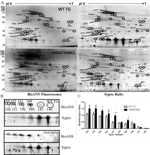
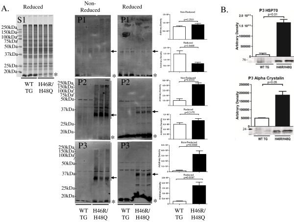
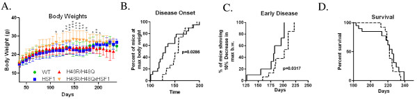
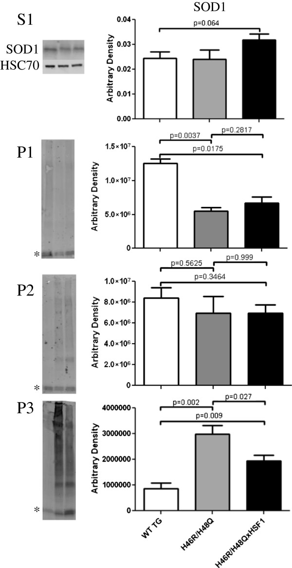
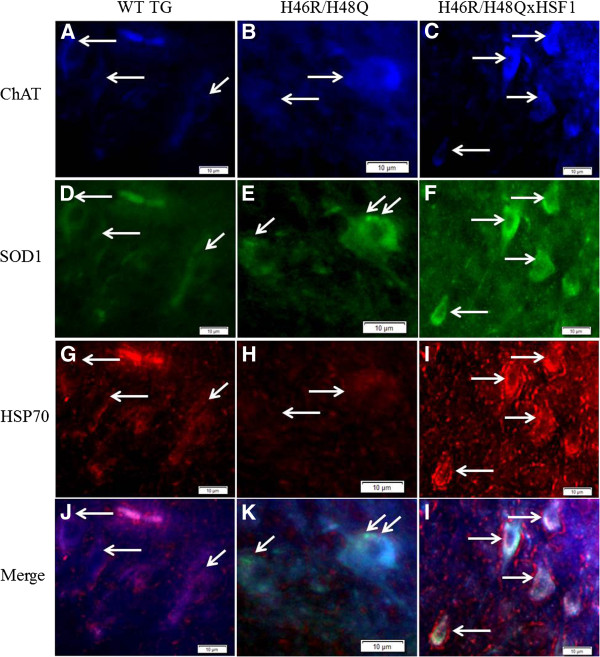
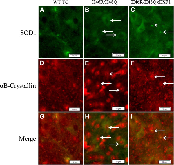
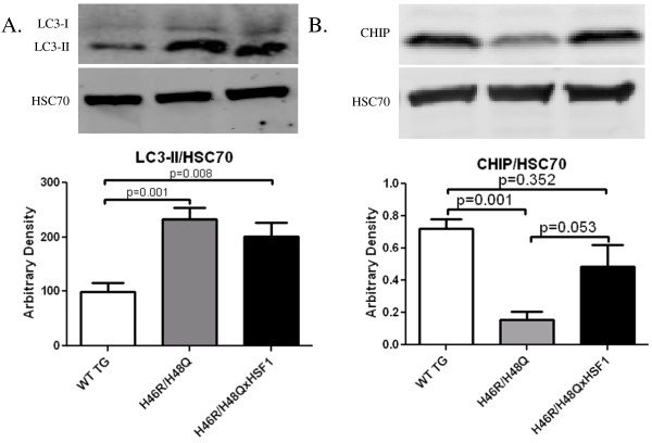
Results: Covalent bisANS labeling of spinal cord extracts revealed that alterations in surface hydrophobicity of H46R/H48Q mutations in SOD1 provoke formation of high molecular weight SOD1 species with lowered solubility, likely due to increased exposure of hydrophobic surfaces. BisANS was docked on the H46R/H48Q SOD1 structure at the disordered copper binding and electrostatic loops of mutant SOD1, but not non-mutant WT SOD1. 16 non-SOD1 proteins were also identified that exhibited altered surface hydrophobicity in the H46R/H48Q mutant mouse model of ALS, including proteins involved in energy metabolism, cytoskeleton, signaling, and protein quality control. Heat shock proteins (HSPs) were also enriched in the detergent-insoluble fractions with SOD1. Given that chaperones recognize proteins with exposed hydrophobic surfaces as substrates and the importance of protein homeostasis in ALS, we crossed SOD1 H46R/H48Q mutant mice with mice over-expressing the heat shock factor 1 (HSF1) transcription factor. Here we showed that HSF1 over-expression in H46R/H48Q ALS mice enhanced proteostasis as evidenced by increased expression of HSPs in motor neurons and astrocytes and increased solubility of mutant SOD1. HSF1 over-expression significantly reduced body weight loss, delayed ALS disease onset, decreases cases of early disease, and increased survival for the 25th percentile in an H46R/H48Q SOD1 background. HSF1 overexpression did not affect macroautophagy in the ALS background, but was associated with maintenance of carboxyl terminus of Hsp70 interacting protein (CHIP) expression which declined in H46R/H48Q mice.

Conclusion: Our results uncover the potential importance of changes in protein surface hydrophobicity of SOD1 and other non-SOD1 proteins in ALS, and how strategies that activate HSF1 are valid therapies for ALS and other age-associated proteinopathies.


Figures

Figure 1
Figure 1
Altered surface hydrophobicity of mutant SOD1 and non-SOD1 proteins in the spinal cords of symptomatic ALS mice. A) Representative 2D gels of wild type transgenic human SOD1 (WT TG, n = 8) and H46R/H48Q (n = 12) with molecular weights (left axis) and isoelectric points (pI, upper axis). Spots that significantly differed from WT TG in hydrophobic ratio are circled and annotated based on the gene names of their accession numbers identified by MALDI-TOF mass spectrometry. B) Enhanced region of 2D gels containing WT SOD1 and H46R/H48Q mutant SOD1 proteins. BisANS fluorescence and corresponding total protein stained with Sypro Ruby are shown. Quantitated SOD1 spots are shown with numbered ellipses and correspond to the quantitated hydrophobic ratio shown in C). Bars represent the mean hydrophobic ratio +/- standard deviation of 10- 8 mice per group. *p<0.05, **p<0.01 by one-way ANOVA.
Figure 2
Figure 2
BisANS docking with SOD1. BisANS (blue) docked with WT human SOD1 [2C9V] and H46R/H48Q [3GQP]. The zinc loop (aa49-84) is colored green, and electrostatic loop (aa121-144) is colored red. Amino acids 46 and 48 are colored magenta, copper ions (orange), and zinc ions (green). Binding energies are given for bisANS binding sites in kcal/mol. H46R/H48Q possess a binding site for bisANS in the metal binding region, whereas holo WT dimeric SOD1 does not.
Figure 3
Figure 3
Mutant SOD1 and chaperones co-fractionate in the detergent insoluble fractions. Spinal cord extracts were subjected to differential detergent extraction and A) the soluble fraction was electrophoresed under reducing and denaturing conditions and stained with Coomassie (S1) and P1-P3 fractions were immunoblotted for SOD1 under non-reducing or reducing conditions (P1-P3). Asterisks indicate the position of the monomeric SOD1. High molecular weight reactivity to the SOD1 antibody was detected in the H46R/H48Q extracts but not WT TG (indicated by vertical bar). High molecular weight reactivity to SOD1 diminished with addition of a reducing agent, however a band corresponding to dimeric SOD1 persisted and is indicated by an arrow. B) Inducible heat shock protein 70 and αB-crystallin co-fractionated with mutant SOD1 in the detergent insoluble fractions of spinal cord. *p = 0.05, **p = 0.01, Bars represent the mean of 6 mice +/- SD.
Figure 4
Figure 4
Expression of HSF1 and distribution of HSPs in spinal cord. A) Western blot of HSF1 in spinal cord homogenates from normal and symptomatic control mice normalized with HSC70, demonstrating the over-expression of HSF1 in H46R/H48QxHSF1 mice compared to H46R/H48Q littermates. B) Spinal cords were extracted with detergents of increasing ionic strength as described in the methods. S1 and P1-3 extracts were immunoblotted for inducible HSP70 (B) or αB-crystallin (C). Asterisks are given at the position of the expected monomeric protein. p is indicated by brackets, bars represent the mean of 6 mice +/- SD.
Figure 5
Figure 5
Effect of HSF1 over-expression on body weight loss and healthspan of H46R/H48Q mice. H46R/H48Q mice (Solid line) n = 20 and H46R/H48QxHSF1 (dashed line) n = 19. A) Beginning at 3 weeks of age, weekly averages of body weight are plotted for the survival cohort for WT (green circles), HSF1 (blue squares), H46R/H48Q (red triangles), and H46R/H48QxHSF1 (gold triangles). Asterisks indicate significant differences in H46R/H48QxHSF1 vs. H46R/H48Q at the *p<0.05, **p<0.01, and ***p<0.001 level. B) Disease onset was determined as the time animals reached their maximum bodyweight. C) Early disease was defined as a drop of 10% of the mouse maximal weight. D) Survival was defined as the point at which animals could not right themselves within 30s after being placed on their side.
Figure 6
Figure 6
Effect of HSF1 over-expression on mutant SOD1 solubility. SOD1 in the soluble S1 and detergent soluble fractions P1-P3. Asterisks are given at the position of the expected monomeric SOD1. Bars represent an n = 6 +/- SD.
Figure 7
Figure 7
Anterior horn region of lumbar spinal cord sections from mice at 220 days. Motor neuron choline acetyltransferase (ChAT) positive cells (A-C, arrows) co-localized with SOD1 positive staining (D-F, arrows). H46R/H48Q mice exhibited small intracellular peri-nuclear SOD1 reactive punctae (E, arrows), while H46R/H48QxHSF1 tissues had a more even intracellular distribution of SOD1 staining (F). This corresponded with differences in HSP70 distribution to SOD1 reactive punctae in H46R/H48Q mice (H) while in contrast, co-localization with ChAT reactive cells in H46R/H48QxHSF1 mice was stronger (I). Scale bar represents 10 μm.
Figure 8
Figure 8
Anterior horn region of lumbar spinal cord sections from mice at 220 days. SOD1 staining (A-C) and small SOD1 positive punctae in H46R/H48Q vs. H46R/H48QxHSF1 tissues (C, arrows) co-localized diffusely with αB-crystallin throughout WT TG and H46R/H48QxHSF1 tissues (D,F). Strikingly, αB-crystallin staining appeared more punctate and localized to cell nuclei of SOD1 expressing cells (E,H, arrows and G-I) in H46R/H48Q tissues. Scale bar represents 10μm.
Figure 9
Figure 9
Effects of HSF1 over-expression on measures of protein quality control in ALS. Whole spinal cords were homogenized in 2%SDS and immunoblotted for A) LC3-II or B) CHIP and normalized with Hsc70. Bars represent an n = 6 +/- SD.

Similar articles

Cited by

References

    1. Rosen DR, Siddique T, Patterson D, Figlewicz DA, Sapp P, Hentati A, Donaldson D, Goto J, O'Regan JP, Deng HX. et al.Mutations in Cu/Zn superoxide dismutase gene are associated with familial amyotrophic lateral sclerosis. Nature. 1993;8:59–62. doi: 10.1038/362059a0. - DOI - PubMed
    1. Kabashi E, Valdmanis PN, Dion P, Spiegelman D, McConkey BJ, Vande Velde C, Bouchard JP, Lacomblez L, Pochigaeva K, Salachas F. et al.TARDBP mutations in individuals with sporadic and familial amyotrophic lateral sclerosis. Nat Genet. 2008;8:572–574. doi: 10.1038/ng.132. - DOI - PubMed
    1. Turner BJ, Talbot K. Transgenics, toxicity and therapeutics in rodent models of mutant SOD1-mediated familial ALS. Prog Neurobiol. 2008;8:94–134. doi: 10.1016/j.pneurobio.2008.01.001. - DOI - PubMed
    1. Valentine JS, Doucette PA, Zittin Potter S. Copper-zinc superoxide dismutase and amyotrophic lateral sclerosis. Annual review of biochemistry. 2005;8:563–593. doi: 10.1146/annurev.biochem.72.121801.161647. - DOI - PubMed
    1. Lelie HL, Liba A, Bourassa MW, Chattopadhyay M, Chan PK, Gralla EB, Miller LM, Borchelt DR, Valentine JS, Whitelegge JP. Copper and zinc metallation status of copper-zinc superoxide dismutase from amyotrophic lateral sclerosis transgenic mice. The Journal of biological chemistry. 2011;8:2795–2806. doi: 10.1074/jbc.M110.186999. - DOI - PMC - PubMed

Publication types

MeSH terms