. 2014 Jan 2;505(7481):97-102.
doi: 10.1038/nature12681. Epub 2013 Nov 20.

Divergent angiocrine signals from vascular niche balance liver regeneration and fibrosis

Affiliations

Divergent angiocrine signals from vascular niche balance liver regeneration and fibrosis

Bi-Sen Ding et al. Nature. .

Abstract

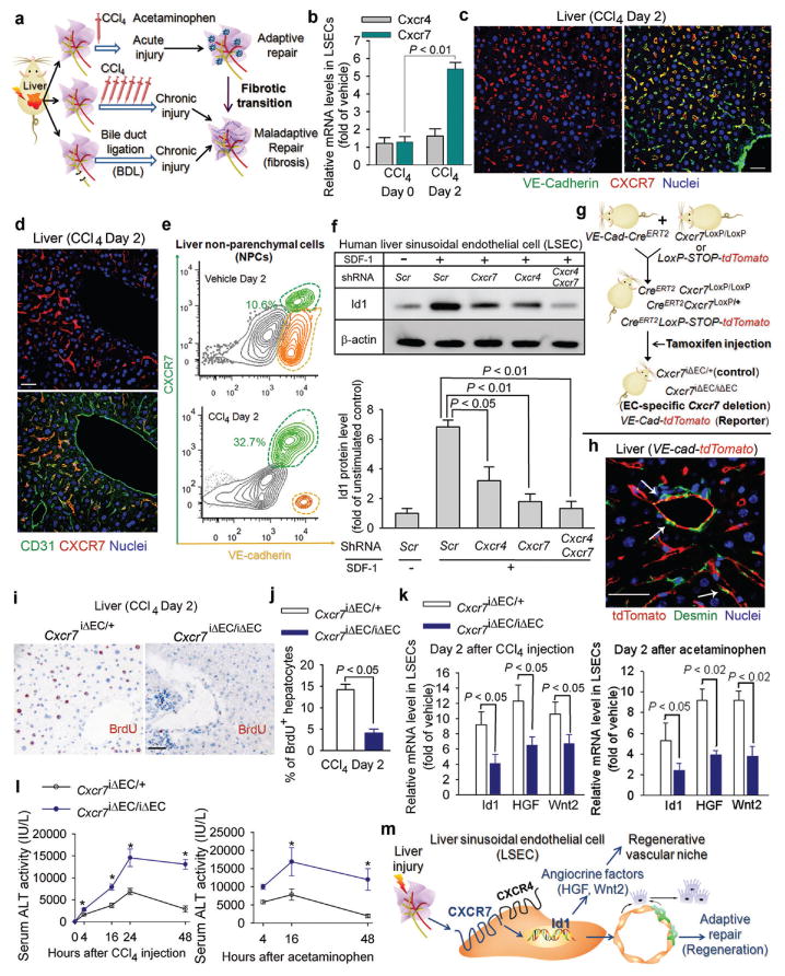
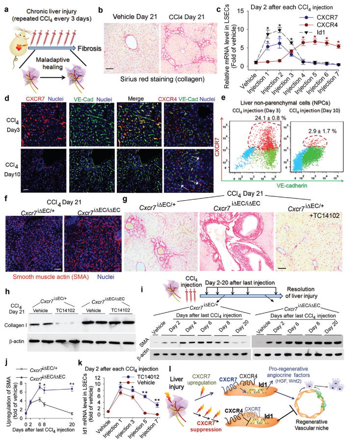
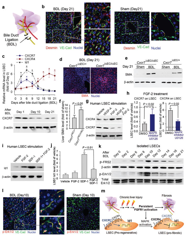
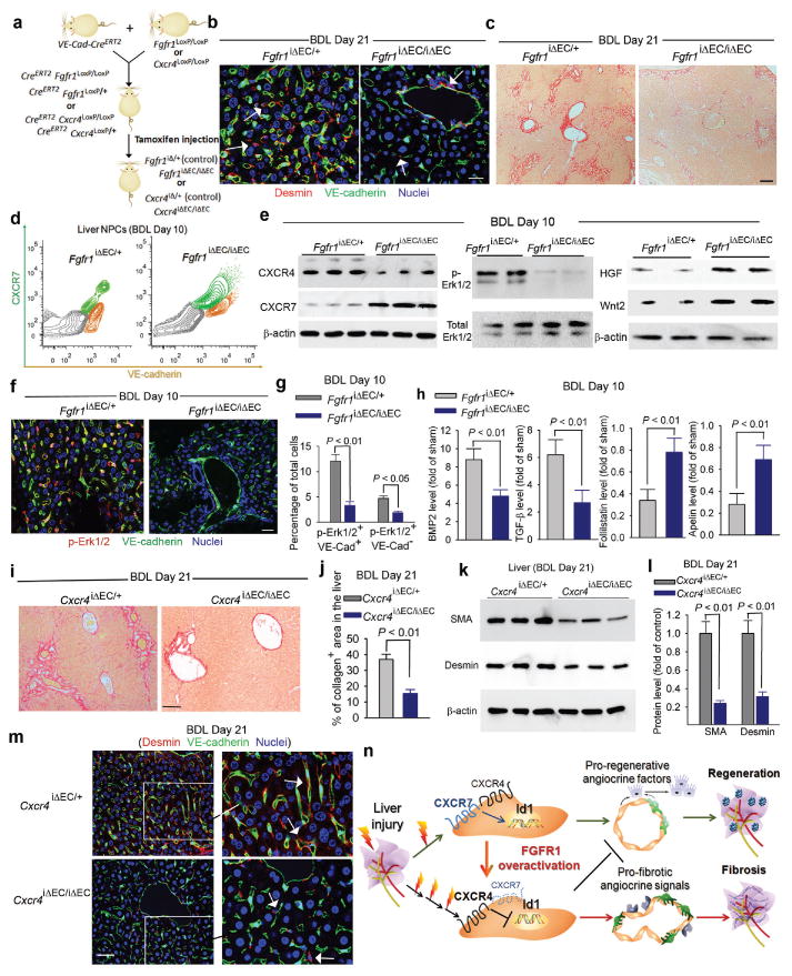
Chemical or traumatic damage to the liver is frequently associated with aberrant healing (fibrosis) that overrides liver regeneration. The mechanism by which hepatic niche cells differentially modulate regeneration and fibrosis during liver repair remains to be defined. Hepatic vascular niche predominantly represented by liver sinusoidal endothelial cells deploys paracrine trophogens, known as angiocrine factors, to stimulate regeneration. Nevertheless, it is not known how pro-regenerative angiocrine signals from liver sinusoidal endothelial cells is subverted to promote fibrosis. Here, by combining an inducible endothelial-cell-specific mouse gene deletion strategy and complementary models of acute and chronic liver injury, we show that divergent angiocrine signals from liver sinusoidal endothelial cells stimulate regeneration after immediate injury and provoke fibrosis after chronic insult. The pro-fibrotic transition of vascular niche results from differential expression of stromal-derived factor-1 receptors, CXCR7 and CXCR4 (refs 18, 19, 20, 21), in liver sinusoidal endothelial cells. After acute injury, CXCR7 upregulation in liver sinusoidal endothelial cells acts with CXCR4 to induce transcription factor Id1, deploying pro-regenerative angiocrine factors and triggering regeneration. Inducible deletion of Cxcr7 in sinusoidal endothelial cells (Cxcr7(iΔEC/iΔEC)) from the adult mouse liver impaired liver regeneration by diminishing Id1-mediated production of angiocrine factors. By contrast, after chronic injury inflicted by iterative hepatotoxin (carbon tetrachloride) injection and bile duct ligation, constitutive FGFR1 signalling in liver sinusoidal endothelial cells counterbalanced CXCR7-dependent pro-regenerative response and augmented CXCR4 expression. This predominance of CXCR4 over CXCR7 expression shifted angiocrine response of liver sinusoidal endothelial cells, stimulating proliferation of desmin(+) hepatic stellate-like cells and enforcing a pro-fibrotic vascular niche. Endothelial-cell-specific ablation of either Fgfr1 (Fgfr1(iΔEC/iΔEC)) or Cxcr4 (Cxcr4(iΔEC/iΔEC)) in mice restored the pro-regenerative pathway and prevented FGFR1-mediated maladaptive subversion of angiocrine factors. Similarly, selective CXCR7 activation in liver sinusoidal endothelial cells abrogated fibrogenesis. Thus, we demonstrate that in response to liver injury, differential recruitment of pro-regenerative CXCR7-Id1 versus pro-fibrotic FGFR1-CXCR4 angiocrine pathways in vascular niche balances regeneration and fibrosis. These results provide a therapeutic roadmap to achieve hepatic regeneration without provoking fibrosis.


Figures

Figure 1
Figure 1. After acute liver injury, upregulation of stromal derived factor (SDF)-1 receptor CXCR7 in liversinusoidal endothelial cells (LSECs) elicits angiocrine-mediated regeneration
a) Liver injury models for studying the maladaptive transition of pro-regenerative LSEC function to a pro-fibrotic vascular niche. b-e) CXCR7 is specifically upregulated on VE-cadherin+CD31+LSECs after acute chemical injury. After injection of carbon tetrachloride(CCl4), CXCR7 and CXCR4 were determined in isolated LSECs (b), liver sections (c, d), and non-parenchymal cells (NPCs) (e). CXCR7 was expressed on LSECs but not large vessels;N=5. Scale bar = 50 μm in Figure 1, all data hereafter are presented as mean ± standard error of mean (s.e.m.). f) SDF-1 stimulation of human LSECs upregulates inhibitor of DNA binding 1 (Id1), a transcription factor inducing production of pro-regenerative angiocrine factors. Id1 stimulation by SDF-1 in primary human Factor VIII+ LSECs was abrogated by silencing of Cxcr4and Cxcr7 in LSECs; N=5. g, h) Endothelial cell (EC)-specific inducible deletion ofCxcr7(Cxcr7iΔEC/iΔEC) in mice. Mice harboring loxPsites-flanked Cxcr7were crossed with mouse line with EC-specific VE-cadherinpromoter-driven CreERT2(VE-Cad-CreERT2). Specificity of VE-Cad-CreERT2 was validated in reporter mice carrying tdTomato protein following floxed stop codon. Cxcr7 deletion or tdTomato expression in ECs was induced by tamoxifen injection.Cxcr7iΔEC/+ mice served as control. Note the specific expression of tdTomato in ECs but not desmin+stellate-like cells (h, white arrows). i-l) Impaired liver regeneration and enhanced liver injury inCxcr7iΔEC/iΔEC mice after acute liver injury. Cell proliferation was determined by staining for BrdU incorporation (i, j). Expression of Id1 and pro-regenerative angiocrine factors, hepatocyte growth factor (HGF) and Wnt2, in LSECswas measured after CCl4 and acetaminophen administration (k), and serum alanine aminotransferase (ALT) level was assessed to determine the degree of liver injury (l); N=5. m) CXCR7 activation in LSECs triggers Id1-mediated production of pro-regenerative angiocrine fctors. After acute liver injury, CXCR7 cooperates with CXCR4, induces pro-regenerative Id1 pathway in LSECs, and triggers angiocrine-mediated liver regeneration.
Figure 2
Figure 2. Iterative hepatotoxic injury perturbs CXCR7 pro-regenerative pathway in LSECs and forces the generation of a pro-fibrotic vascular niche
a-b) Mouse liver fibrosis is induced by repeated injection of CCl4. Sirius red staining was used to detect collagen in the injured liver. Scale br= 50 μm in Figure 2. c-e) Chronic liver injury suppresses CXCR7 pathway and upregulates CXCR4 expression in LSECs. Quantitative PCR (c), immunostaining (d) and flow cytometry (e) showed the abrogationof CXCR7-Id1 pathway in VE-cadherin (VE-Cad)+ LSECs after chronic CCl4 injury. CXCR4 is expressed in both ECs and non-ECs (white arrow). *, P< 0.05 versus vehicle-treated mice; N=8. f-h) CXCR7 activation in LSECs negates liver fibrosis. The extent of fibrosis was augmented in Cxcr7iΔEC/iΔEC mice, as evidenced by elevated hepatic levels of α-smooth muscle actin (SMA) and collagen I. Notably, CXCR7-selective agonist TC14012 reduced fibrosis in control but not Cxcr7iΔEC/iΔEC mice; N=7. i, j) Impaired resolution of liver injury inCxcr7iΔEC/iΔEC mice. SMA level in the injured liver was tested to assess the resolution of injury (i). Compared to control mice, SMA level in Cxcr7iΔEC/iΔEC mice was enhanced after last CCl4 injection and remained stable afterwards. Collagen I level was similarly assessed (supplementary Figure 13); *, P < 0.05, **, P < 0.01, versus control mice; N=5. k) CXCR7 activation restores Id1 induction in chronically injured LSECs. TC14102 prevented Id1 suppression in LSECs during repeated CCl4 injury. *, P < 0.05, **, P < 0.01, compared to vehicle group; N=7. l) Interference with pro-regenerative CXCR7-Id1 pathway in LSECs causes pro-fibrotic transition of vascular niche. After injury, upregulation of the CXCR7-Id1 pathway in LSECs induces generation of hepatic-active angiocrine factors and stimulates regeneration. Chronic injury perturbs CXCR7-Id1 signaling, counteracting regeneration and provoking fibrogenesis.
Figure 3
Figure 3. Cholestatic liver injury via FGFR1 overactivation in LSECs shifts CXCR7-dependent pro-regenerative response to a CXCR4-dominated pro-fibrotic vascular niche
a, b) Pro-fibrotic transition of LSECs caused by bile duct ligation (BDL)-induced cholestatic injury. After BDL-induced cholestatic liver injury (a), majority of VE-cadherin+ LSECs were covered by perisinusoidal desmin+ fibroblasts (b, inset). By contrast, desmin+ stellate-like cells were sparsely distributed in sham-operated liver. Scale bar = 50 μm in Figure 3. c) BDL suppresses CXCR7-Id1 pathway and upregulates CXCR4 expression in LSECs. Loss of CXCR7 protein in LSECs after BDL is shown at bottom panel; *, P < 0.05, compared with the level at day 0; N=5. d-f) Liver fibrosis caused by BDL is exacerbated inCxcr7iΔEC/iΔECmice. After BDL, Cxcr7iΔEC/iΔEC mice exhibited higher hepatic level of SMA protein than that of control mice; N=4. g-j) FGF-2 via MAP kinase activation favors CXCR4 signaling in LSECs, counteracting CXCR7-Id1 pathway. FGF-2, but not VEGF-A, upregulated CXCR4, suppressed CXCR7, and inhibited SDF-1-dependent Id1 induction in LSECs. This FGF-2-mediated predominance of CXCR4 over CXCR7 was attenuated by MAP kinase (MAPK) inhibitor U0126; N=5. k, l) Activation of FGFR1 and MAPK pathway in LSECs after BDL. There was time-dependent enhancement in phosphorylation/activation of FGFR1 downstream effector FRS2 (p-FRS2) and Erk1/2 (p-Erk1/2) in VE-cadherin+ LSECs after BDL; N=6. m) Constitutive FGFR1 signaling in LSECs via MAPK activation forces a CXCR4-dominated pro-fibrotic vascular niche. During chronic liver injury, FGFR1-mediated aberrant MAPK activation in LSECs upregulates CXCR4 and perturb CXCR7-Id1 pathway. This predominance of FGFR1/CXCR4 activation in LSECs determines the pathological progression from adaptive (pro-regenerative) to maladaptive (pro-fibrotic) liver repair.
Figure 4
Figure 4. FGFR1 activation of CXCR4 in LSECs provokes pro-fibrotic angiocrine signals in liver repair
a) EC-specific inducible deletion ofFgfr1and Cxcr4(Fgfr1iΔEC/iΔECand Cxcr4 iΔEC/iΔEC) in adult mice. b, c) Reduced liver fibrosis in Fgfr1iΔEC/iΔEC mice. Compared to control mice, perisinusoidal enrichment of desmin+ stellate-like cells (b, white arrow) and collagen deposition (c) were diminished in the liver of Fgfr1iΔEC/iΔEC mice after BDL; N=4; Scale bar = 50 μm in Figure 4. d-g) EC-specific deletion of Fgfr1 in mice prevents CXCR4-mediated maladaptive transition after BDL and restores regenerative angiocrine signals. In BDL-injured LSECs of Fgfr1iΔEC/iEC mice, CXCR7 suppression and CXCR4 upregulation (d) and Erk1/2 activation in VE-cadherin+ LSECs (e-g) were reduced. This was accompanied by restored production of hepatic-active angiocrine factors HGF and Wnt2; N = 4. h) Pro-fibrotic production of agiocrine factors in LSECs is reduced in Fgfr1iΔEC/iΔEC mice. BDL instigated divergent production of angiocrine factors in LSECs, including upregulation of factors in BMP andTGF-β pathways and suppression of anti-fibrotic genes such as follistatin and apelin (supplementary Fig. 19). This pro-fibrotic drift of angiocrine factor in LSECs after BDL was mitigated i Fgfr1iΔEC/iΔEC mice; N=4. i-m) Reduction of liver fibrosis in Cxcr4iΔEC/iΔEC mice after BDL. The extent of liver fibrosis after BDL was significantly lower in Cxcr4iΔEC/iΔEC mice than that of control mice, as evidenced by decreased deposition of collagen (i, j),SMA (k, l), and perisinusoidal enrichment of desmin+ stellate-like cells (m, white arrow); N=5. n) Divergent angiocrine signals from LSECs balance liver regeneration and fibrosis. After acute liver injury, activation of CXCR7-Id1 pathway in LSECsstimulates production of hepatic-active angiocrine factors. By contrast, chronic injury causes overt FGFR1 activation in LSECs that perturbs CXCR7-Id1 pathway and favors a CXCR4-driven pro-fibrotic angiocrine response, thereby provoking liver fibrosis. Therefore, in response to injury, differentially primed LSECs deploy divergent angiocrine signals to balance liver regeneration and fibrosis.

Comment in

Similar articles

Cited by

References

    1. Friedman SL, Sheppard D, Duffield JS, Violette S. Therapy for fibrotic diseases: nearing the starting line. Science translational medicine. 2013;5:161sr167. - PubMed
    1. Bataller R, Brenner DA. Liver fibrosis. J Clin Invest. 2005;115:209–218. - PMC - PubMed
    1. Iredale JP. Models of liver fibrosis: exploring the dynamic nature of inflammation and repair i a solid organ. J Clin Invest. 2007;117:539–548. - PMC - PubMed
    1. Gurtner GC, Werner S, Barrandon Y, Longaker MT. Wound repair and regeneration. Nature. 2008;453:314–321. - PubMed
    1. Wynn TA. Common and unique mechanisms regulate fibrosis in various fibroproliferative diseases. J Clin Invest. 2007;117:524–529. - PMC - PubMed

Publication types

MeSH terms