. 2013 Nov 19;14(11):22741-52.
doi: 10.3390/ijms141122741.

Small molecule binding, docking, and characterization of the interaction between Pth1 and peptidyl-tRNA

Affiliations

Small molecule binding, docking, and characterization of the interaction between Pth1 and peptidyl-tRNA

Mary C Hames et al. Int J Mol Sci. .

Abstract

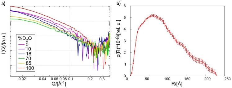
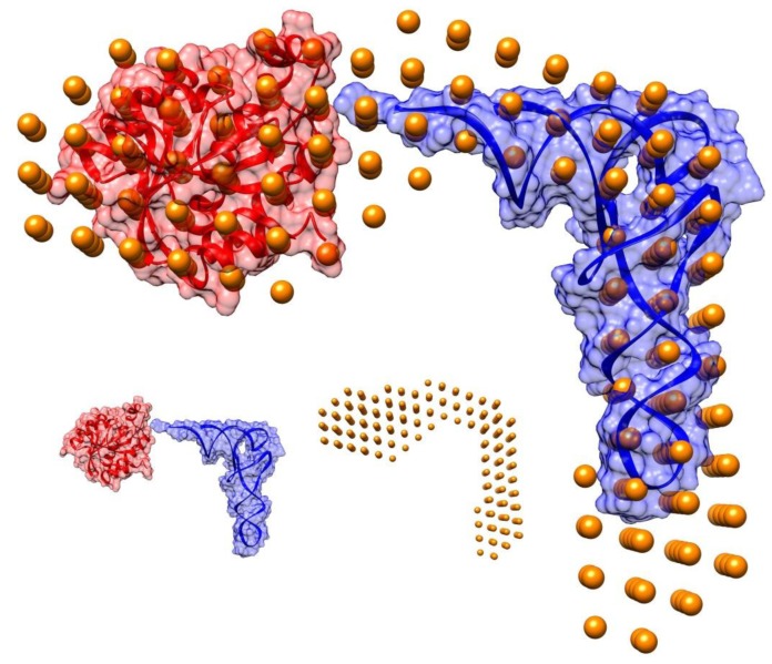
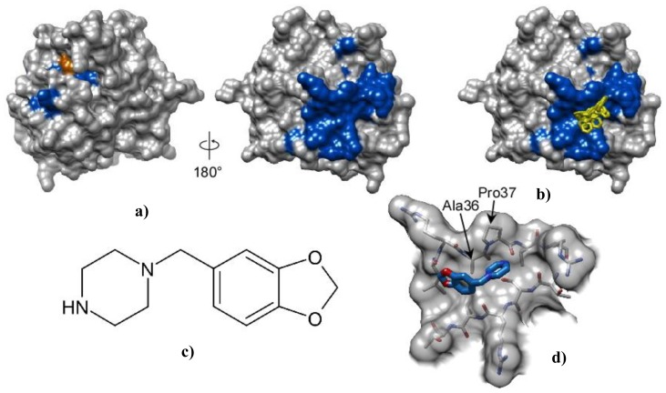
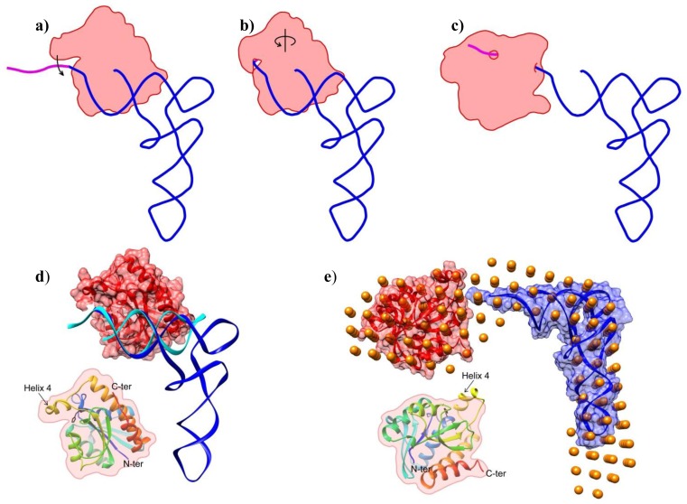
Bacterial Pth1 is essential for viability. Pth1 cleaves the ester bond between the peptide and nucleotide of peptidyl-tRNA generated from aborted translation, expression of mini-genes, and short ORFs. We have determined the shape of the Pth1:peptidyl-tRNA complex using small angle neutron scattering. Binding of piperonylpiperazine, a small molecule constituent of a combinatorial synthetic library common to most compounds with inhibitory activity, was mapped to Pth1 via NMR spectroscopy. We also report computational docking results, modeling piperonylpiperazine binding based on chemical shift perturbation mapping. Overall these studies promote Pth1 as a novel antibiotic target, contribute to understanding how Pth1 interacts with its substrate, advance the current model for cleavage, and demonstrate feasibility of small molecule inhibition.


Figures

Figure 1
Figure 1
Small Angle Neutron Scattering. (a) Scattering curves for Pth1H20R:peptidyl-tRNA complex from contrast series measurements taken at buffer D2O concentrations of 0%, 10%, 18%, 70%, 85%, and 100%; (b) Pairwise distance distribution function of scattering data from complex in 100% D2O generated in GNOM [25].
Figure 2
Figure 2
Model of Pth1:peptidyl-tRNA Complex. The overall shape of the Pth1H20R:peptidyl-tRNA complex is shown in gold spheres. E. coli Pth1 (PDBID: 2PTH) and tRNAPhe (PDBID:1EHZ) were fit into the mass density. Pictured in the inset (lower right) are the individual components: tRNAPhe in blue, Pth1 in red, and the calculated shape in gold spheres.
Figure 3
Figure 3
Interaction and docking of E. coli Pth1 with piperonylpiperazine. (a) Surface representation of E. coli Pth1 (PDBID:2PTH) shown with catalytically important His20 in orange. From NMR data, residues with 1H–15N resonances affected by interaction with piperonylpiperazine are in blue; (b) Docking: The six lowest energy orientations of piperonylpiperazine are shown in yellow; (c) Structure of piperonylpiperazine; (d) An enlarged view of the piperonylpiperazine binding site.
Figure 4
Figure 4
Model of Pth1 Interaction with peptidyl-tRNA. (ac) Cartoon representation of the Pth1 (red) interaction model with peptidyl-tRNA (blue and magenta). (a) After substrate recognition; (b) helix 4 clamps the peptide portion (magenta) and CCA terminus of the substrate in the binding channel; (c) followed by the enzymatic reaction and release of products or just release of the nucleotide as observed in the SANS model; (de) Available high and low resolution structures of Pth1 and peptidyl-tRNA on which the model of interaction was built; (d) Crystal structures of the complex between Pth1 (PDBID:2PTH, red surface) and the TΨC loop of tRNA (PDBID:3VJR, cyan) with tRNAPhe(PDBID:1EHZ, blue) superimposed; (e) SANS model (orange beads) of the interaction presented here with the same coloring as in (d); Insets show the orientation of Pth1. In black, His20 is the only side chain shown.

Similar articles

Cited by

References

    1. Jorgensen F., Kurland C.G. Processivity errors of gene expression in Escherichia coli. J. Mol. Biol. 1990;215:511–521. - PubMed
    1. Manley J.L. Synthesis and degradation of termination and premature-termination fragments of beta-galactosidase in vitro and in vivo. J. Mol. Biol. 1978;125:407–432. - PubMed
    1. Kurland C.G., Ehrenberg M. Constraints on the accuracy of messenger RNA movement. Q. Rev. Biophys. 1985;18:423–450. - PubMed
    1. Heurgue-Hamard V., Karimi R., Mora L., MacDougall J., Leboeuf C., Grentzmann G., Ehrenberg M., Buckingham R.H. Ribosome release factor RF4 and termination factor RF3 are involved in dissociation of peptidyl-tRNA from the ribosome. EMBO J. 1998;17:808–816. - PMC - PubMed
    1. Karimi R., Pavlov M.Y., Heurgue-Hamard V., Buckingham R.H., Ehrenberg M. Initiation factors IF1 and IF2 synergistically remove peptidyl-tRNAs with short polypeptides from the P-site of translating Escherichia coli ribosomes. J. Mol. Biol. 1998;281:241–252. - PubMed

Publication types

MeSH terms

LinkOut - more resources