. 2013 Nov 21:10:143.
doi: 10.1186/1742-4690-10-143.

The S40 residue in HIV-1 Gag p6 impacts local and distal budding determinants, revealing additional late domain activities

Affiliations

The S40 residue in HIV-1 Gag p6 impacts local and distal budding determinants, revealing additional late domain activities

Susan M Watanabe et al. Retrovirology. .

Abstract

Background: HIV-1 budding is directed primarily by two motifs in Gag p6 designated as late domain-1 and -2 that recruit ESCRT machinery by binding Tsg101 and Alix, respectively, and by poorly characterized determinants in the capsid (CA) domain. Here, we report that a conserved Gag p6 residue, S40, impacts budding mediated by all of these determinants.

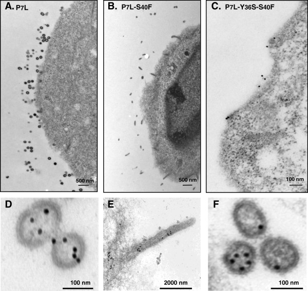
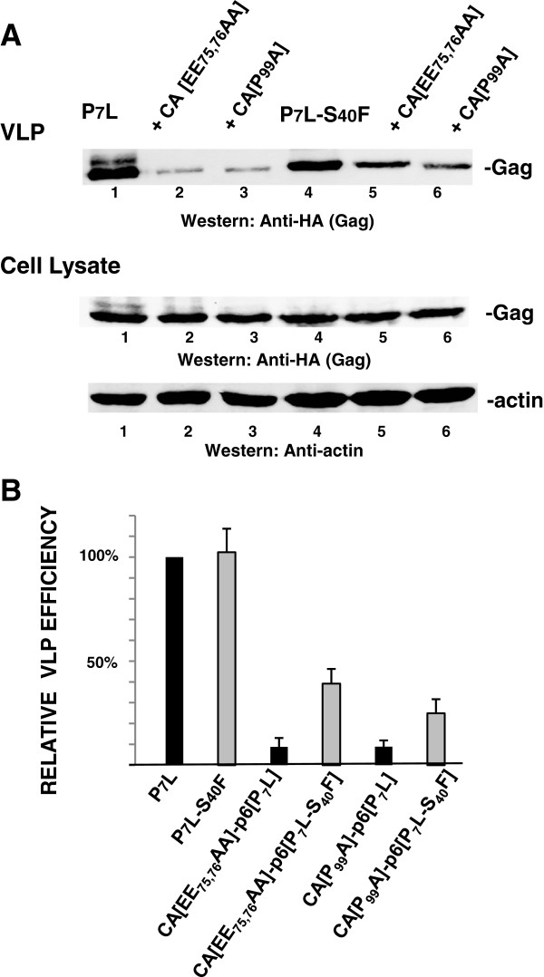
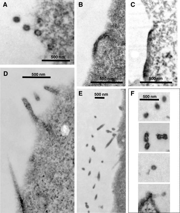
Results: Whereas budding normally results in formation of single spherical particles ~100 nm in diameter and containing a characteristic electron-dense conical core, the substitution of Phe for S40, a change that does not alter the amino acids encoded in the overlapping pol reading frame, resulted in defective CA-SP1 cleavage, formation of strings of tethered particles or filopodia-like membrane protrusions containing Gag, and diminished infectious particle formation. The S40F-mediated release defects were exacerbated when the viral-encoded protease (PR) was inactivated or when L domain-1 function was disrupted or when budding was almost completely obliterated by the disruption of both L domain-1 and -2. S40F mutation also resulted in stronger Gag-Alix interaction, as detected by yeast 2-hybrid assay. Reducing Alix binding by mutational disruption of contact residues restored single particle release, implicating the perturbed Gag-Alix interaction in the aberrant budding events. Interestingly, introduction of S40F partially rescued the negative effects on budding of CA NTD mutations EE75,76AA and P99A, which both prevent membrane curvature and therefore block budding at an early stage.

Conclusions: The results indicate that the S40 residue is a novel determinant of HIV-1 egress that is most likely involved in regulation of a critical assembly event required for budding in the Tsg101-, Alix-, Nedd4- and CA N-terminal domain affected pathways.


Figures

Figure 1
Figure 1
Mutation of S40 to Phe, which does not alter the amino acid sequence in the overlapping pol reading frame, nevertheless inhibits CA maturation, alters budding, and reduces viral infectivity. Panels A1-A3, Western blot analysis of pNL4-3ΔEnv WT, S40A and S40F constructs which encode active protease. VLPs and cell lysates from COS-1 cells transfected with pNL4-3ΔEnv-WT (lane 1), pNL4-3ΔEnv-S40A (lane 2) or pNL4-3ΔEnv-S40F (lane 3) were analyzed by Western blotting. The values in the figure indicate the ratio of p25/p24 normalized to wild type. The relative VLP efficiencies [VLP/(VLP + Gag from cell lysate)] were determined as described in Methods. Panel B, Electron microscopy of particles associated with cells transfected with pNL4-3ΔEnv-WT (panel B1), or S40F (panel B2). Immature particles (open arrows), particles with aberrant cores (gray arrows), and mature particles with conical cones (solid arrows) were released from pNL4-3ΔEnv-transfected cells. Panel C, Quantitative analysis. 25 WT or 100 S40F VLPs (n =2) were counted and the frequency of each type of particle was determined. Panel D, Viral particle infectivity, determined by MAGI assay, done in triplicate.
Figure 2
Figure 2
The budding defect resulting from S40F mutation becomes more apparent when PR is inactive. COS-1 cells were transfected with plasmids expressing HA-tagged HIV-1 Gag-WT (panels A and B) or Gag-S40F (panels C and D). Cells were prepared for examination by electron microscopy as described in Methods. Panels A and C, solid arrows, immature particles; open arrows, extended or tethered particles. Bars in panels A and C measure 500 nm; bars in panel B and D measure 100 nm. The relative frequencies of typical spherical particles and filopodia-like particles are summarized (panel E).
Figure 3
Figure 3
Disrupting L domain-1 increases the effect of the S40F mutation on budding. COS-1 cells were transfected with plasmids expressing HA-tagged HIV-1 Gag-P7L (panels A1 and A2) or P7L-S40F (panels B1 and B2) and prepared for examination by electron microscopy as described in Methods. Panels A2, and B2: Samples were labeled with primary rabbit anti-CA antibody that was tagged with 15 nm gold-conjugated mouse anti-rabbit secondary antibody (bars in panels A1 and A2 measure 100 nm; bars in panels B1 and B2 measure 500 nm). Panel C, Quantitative analysis of particle morphology. 200 gold-tagged VLPs were counted and the frequency of spherical versus extended structures were determined (n =2). Panel D, Western Analysis. VLPs and cell lysates from the COS-1 cells transfected with HA-tagged HIV-1 Gag-P7L (lane 1), P7L-S40F (lane 2) were analyzed by Western blotting and gag related proteins identified by monoclonal HA antibody. Panel E, the relative VLP release efficiencies [VLP/(VLP + Gag from cell lysate)] were determined as described in Methods.
Figure 4
Figure 4
Gag p6-S40 mutations increase binding to Alix in the yeast 2-hybrid assay. Panel A, Qualitative colony assay. Co-transformed plasmids encoding protein pairs were tested for interaction under permissive and selective conditions using double drop-out media (DDO, Trp and Leu) and triple drop-out media (TDO, Trp, Leu, His), respectively. All transformed cells grew on the DDO media, indicating that plasmid pairs were expressed; only cells expressing interacting pairs grew on the TDO media. Panel B, Quantitative liquid assay. Beta-galactosidase assays were used to measure the relative strength of the S40 mutants plus Alix interaction. Beta-galactosidase units were normalized to P7L paired with Alix. Tsg101 plus P7L and Alix plus P7L-Y36S are negative controls. The S40A and S40F results represent five independent clones tested in 5 independent assays. Panel C, The beta-galactosidase signal is undetectable when the S40 mutants are paired with Alix F676D. WT Gag, P7L-S40A and P7L-S40F were each paired with wild type Alix or its F676D mutant which does not bind to Gag. Alix paired with Y36S serves as the negative control. Two independent clones were run in duplicate for each pairing. All values were normalized to WT paired with Alix.
Figure 5
Figure 5
Disrupting Alix interaction with S40F restores spherical particle formation. COS-1 cells were transfected with plasmids expressing HA-tagged -P7L (panels A and D), P7L-S40F (panels B and E), or P7L-Y36S-S40F (panels C and F). Panels C-F, cells were prepared for examination by immunoelectron microscopy as described in Methods. Bars in panels A, and B measure 500 nm; bars in panels C, D and F measure 100 nm; bar in panel E measures 2000 nm.
Figure 6
Figure 6
The S40F mutation increases the disruption of virus release due to nonfunctional L domains-1 and −2. Panel A, COS-1 cells transfected with DNA encoding the indicated constructs were analyzed by Western blotting after preparation of VLPs and cell lysates. Panel B, Quantitative analysis of VLP release efficiency [VLP/(VLP + Gag from cell lysate)] were determined as described in Methods (n = 7; 3 independent constructs of P7L-S40F-Gag).
Figure 7
Figure 7
The S40F mutation partially rescues the block to VLP release efficiency imposed by mutations in the CA NTD. Panel A, COS-1 cells transfected with DNA encoding the indicated constructs were analyzed by Western blotting after preparation of VLPs and cell lysates. Panel B, Quantitative analysis of relative VLP release efficiency [VLP/(VLP + Gag from cell lysate)] were determined as described in Methods (n = 3).
Figure 8
Figure 8
The S40F mutation suppresses the block to budding imposed by CA[EE75,76AA] and CA[P99A]. The samples described in the legend to Figure 7 were analyzed by electron microscopy. F. CA[P99A]-p6[P7L-S40F] Panel A, P7L; panel B, CA [EE75,76AA]-p6[P7L]; panel C, CA[P99A]-p6[P7L]; panel D, P7L-S40F; panel E, CA[EE75,76AA]-p6[P7L-S40F]; and panel F, CA[P99A]-p6[P7L-S40F].

Similar articles

Cited by

References

    1. Gottlinger HG. The HIV-1 assembly machinery. AIDS. 2001;10(Suppl 5):S13–20. - PubMed
    1. Lalonde MS, Sundquist WI. How HIV finds the door. Proc Natl Acad Sci USA. 2012;10:18631–18632. doi: 10.1073/pnas.1215940109. - DOI - PMC - PubMed
    1. Votteler J, Sundquist WI. Virus Budding and the ESCRT Pathway. Cell Host Microbe. 2013;10:232–241. doi: 10.1016/j.chom.2013.08.012. - DOI - PMC - PubMed
    1. Meng B, Lever AM. Wrapping up the bad news: HIV assembly and release. Retrovirology. 2013;10:5. doi: 10.1186/1742-4690-10-5. - DOI - PMC - PubMed
    1. Garrus JE, Von Schwedler UK, Pornillos OW, Morham SG, Zavitz KH, Wang HE, Wettstein DA, Stray KM, Cote M, Rich RL, Myszka DG, Sundquist WI. Tsg101 and the vacuolar protein sorting pathway are essential for HIV-1 budding. Cell Dev Biol. 2001;10:55–65. - PubMed

Publication types

MeSH terms

Substances