Atypical cerebellar slump syndrome and external hydrocephalus following craniocervical decompression for Chiari I malformation: case report
- PMID: 24257499
- PMCID: PMC4533469
- DOI: 10.2176/nmc.cr2013-0066
Atypical cerebellar slump syndrome and external hydrocephalus following craniocervical decompression for Chiari I malformation: case report
Abstract
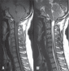
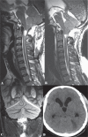
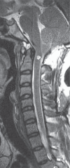
Symptomatic cerebellar slump (CS) and external hydrocephalus (EH) are amongst the rarer complications of foramen magnum decompression (FMD) for Chiari I malformation (CM). CS typically presents with delayed onset headache related to dural traction or with neurological deficit offsetting the benefit of FMD. EH, consisting of ventriculomegaly along with subdural fluid collection(s) (SFCs), has been related to cerebrospinal fluid egress from a tiny breach in an otherwise intact arachnoid. We describe the case of a 21-year-old man with CM and syringomyelia who presented with impaired gag, spastic quadriparesis, and raised intracranial pressure 1 week following an uneventful FMD during which the arachnoid had been widely fenestrated. Magnetic resonance imaging (MRI) showed an infratentorial SFC, dilated aqueduct and triventriculomegaly, features of CS, and a residual but resolving syrinx. His symptoms resolved following a high pressure ventriculo-peritoneal shunt. At a 6-month follow-up visit, he was asymptomatic and demonstrated partial resolution of the syrinx, with no recurrence of the SFC. The unusual features in the clinical course of this patient were an atypical CS syndrome presenting with concomitantly resolving syringomyelia, and the development of EH after a wide arachnoidal fenestration. This is the first case in indexed literature describing such a combination of unusual postoperative complications of a FMD. A hypothesis is presented to explain the clinico-radiological findings of the case.
Conflict of interest statement
The authors have not received any support, in the form of grant, from any source for preparation of this article. Neither do the authors nor does the institute have any personal or institutional financial interest in drugs, materials, or devices described in their submissions.
This article has not been published before and has not been submitted for publication to any other journal in part or full. It has not been presented in any meeting/conference. The authors concur with this submission and there is no conflict of interest arising from this article.
Figures


Similar articles
-
Subdural Fluid Collection and Hydrocephalus After Foramen Magnum Decompression for Chiari Malformation Type I: Management Algorithm of a Rare Complication.World Neurosurg. 2017 Oct;106:1057.e9-1057.e15. doi: 10.1016/j.wneu.2017.07.112. Epub 2017 Jul 25. World Neurosurg. 2017. PMID: 28754644 Review.
-
Management of infratentorial subdural hygroma complicating foramen magnum decompression: a report of three cases.Acta Neurochir (Wien). 2011 May;153(5):1123-8. doi: 10.1007/s00701-010-0920-2. Epub 2011 Jan 22. Acta Neurochir (Wien). 2011. PMID: 21258949
-
Subacute subdural hygroma and presyrinx formation after foramen magnum decompression with duraplasty for Chiari type 1 malformation.Neurol Med Chir (Tokyo). 2011;51(5):389-93. doi: 10.2176/nmc.51.389. Neurol Med Chir (Tokyo). 2011. PMID: 21613769
-
Shifting subdural fluid collection and hydrocephalus after foramen magnum decompression for basilar invagination.Br J Neurosurg. 2020 Jun;34(3):321-323. doi: 10.1080/02688697.2020.1716941. Epub 2020 Jan 24. Br J Neurosurg. 2020. PMID: 31975622
-
Acute obstructive hydrocephalus associated with infratentorial subdural hygromas complicating Chiari malformation Type I decompression. Report of two cases and literature review.J Neurosurg. 2005 Oct;103(4):752-5. doi: 10.3171/jns.2005.103.4.0752. J Neurosurg. 2005. PMID: 16266060 Review.
Cited by
-
Retrocerebellar arachnoid cyst resulting in syringomyelia in a patient without tonsillar herniation: successful surgical treatment with reconstruction of CSF flow in the foramen magnum region.Neurosurg Rev. 2016 Apr;39(2):341-6; discussion 347. doi: 10.1007/s10143-015-0680-9. Epub 2016 Jan 4. Neurosurg Rev. 2016. PMID: 26728365
-
Neonatal Ventricular Reservoir Implantation for Hydrocephalus Management in Chiari III Malformations: A Case Report.Cureus. 2024 Mar 10;16(3):e55896. doi: 10.7759/cureus.55896. eCollection 2024 Mar. Cureus. 2024. PMID: 38595901 Free PMC article.
References
-
- Menezes AH: Chiari I malformations and hydromyelia—complications. Pediatr Neurosurg 17: 146– 154, 1991–1992. - PubMed
-
- Perrini P, Benedetto N, Tenenbaum R, Di Lorenzo N: Extra-arachnoidal cranio-cervical decompression for syringomyelia associated with Chiari I malformation in adults: technique assessment. Acta Neurochir (Wien) 149: 1015– 1022; discussion 1022–1023, 2007. - PubMed
-
- Furtado SV, Visvanathan K, Reddy K, Hegde AS: Pseudotumor cerebri: as a cause for early deterioration after Chiari I malformation surgery. Childs Nerv Syst 25: 1007– 1012, 2009. - PubMed
-
- Hirano Y, Sugawara T, Sato Y, Sato K, Omae T, Sasajima T, Mizoi K: Negative pressure pulmonary edema following foramen magnum decompression for Chiari malformation type I. Neurol Med Chir (Tokyo) 48: 137– 139, 2008. - PubMed
-
- Vergani F, Nicholson C, Jenkins A: Tethering of the cervicomedullary junction with central cord oedema after foramen magnum decompression for Chiari malformation. Br J Neurosurg 25: 327– 329, 2011. - PubMed
Publication types
MeSH terms
LinkOut - more resources
Full Text Sources
Other Literature Sources
Medical

